3D Lily Section Section
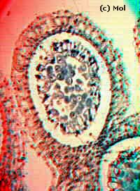
This section of a lily bud was created by taking two pictures at the microscope.
The slide was moved along the x-axis and the instrument was refocused between
shots to obtain finer detail in the middle of the specimen. You can
see how the specimen consists of processes around the outside which are at
on a different plane to those in the middle.
The two 'source images were combined using Double Vision software.
Comments and errors to The Editor
(c) www.microscopy-uk.net 1995-96 UK.
© Onview.net Ltd, Microscopy-UK, and all contributors 1995 onwards. All rights
reserved. Main site is at www.microscopy-uk.org.uk with full mirror at www.microscopy-uk.net.