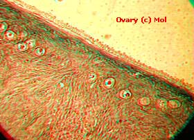
3D Mammalian Ovary

This is a section through part of a mammalian ovary. The tiny rows
of cells are primary follicles. Some of these will ultimately
mature to form Graafian follicles.
The image was created by 'sampling' the source slide into a computer
from an optical microscope by using a camcorder. An image was 'taken', and
then the glass specimen slide was moved left before a second image
was shot. Boths images were then used with Double Vision software to form the 3D representation
you see here.
The depth of the specimen is very shallow so the 3D effect is slight.
Comments and errors to The Editor
(c) www.microscopy-uk.net 1995-96 UK.
© Onview.net Ltd, Microscopy-UK, and all contributors 1995 onwards. All rights
reserved. Main site is at www.microscopy-uk.org.uk with full mirror at www.microscopy-uk.net.

