Human Eyes
Humans and most mammals have Complex Eyes which use a very sophisticated lens system to focus light onto receiving
nerve sensors at the back of the eye. This type of eye cannot sample the outside world at the same fast speed of
the insect's compound eye, and is a lot slower. However, the information gathered is highly detailed.
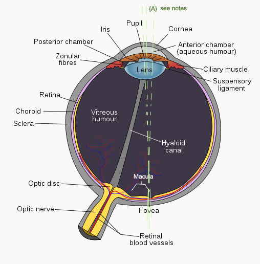
On the right is a diagram of a human eye. The Retina
has two distinct types of light-sensitive nerve cells: rods and cones.
Rods detect monochrome (black-and-white) imagery and are especially well suited to low light levels. There are
no rods at the Fovea or blind spot, and more exist for peripheral vision than central vision. The Fovea region
is highly important in the human eye, as it contains more cones than in other parts of the eye. This allows much
greater detail in our central vision (see A in diagram) where light waves are gathered and focused onto the Fovea
through the centre of the pupil. Thus we receive a sharper picture when we look directly at an object.
Cones enable color vision but they need brighter light to function than rods. Cones come in three types: long-wavelength sensitive (red colour detection), medium-wavelength
sensitive (green colour detection), and short-wavelength
sensitive light (blue
colour detection).
Cone and rod cells connect to nerve fibres of the optic nerve through intermediary cells in the retina. When light
falls on the rods and cones, nerves send electrical impulses into the brain.
Stereo Vision
and IPD The astounding things about human eyes is that they
auto-focus and they also adjust their irises to control light input. They are self-cleaning, and because we have
two of them placed side-by side, they provide us with two separate views of the external world - an attribute which
the brain uses by combining the two images into a single stereoscopic 3D image. This aids us in determining depth
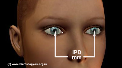
and the relative distances of objects in our field of view. The distance between your two pupils(centres) is called
the Interpupillary distance (IPD). It obviously determines the stereo
separation of the two images which are combined in the brain to produce stereo perception.
Stereoscopic displays and stereoscopic projectors
need to function with some mean value for human eye IPD to ensure we perceive the 3D aspect correctly. In recent
months, many advances have taken place in technology and media content (Avatar Movie) to promote stereoscopic 3D
viewing. Up until recently, very little real study has taken place to determine the IPD in various population and
cultural groups and gender. However, a good mean value has now been established of a distance equal to 63mm.
The following external link details a good paper on IPD research: variation and extrema of human interpupillary
distance by Neil A. Dodgson* University
of Cambridge Computer Laboratory, 15 J. J. Thomson Avenue, Cambridge, UK CB3 0FD
Blind
Spot
Our eyes have a blind spot. This is the area at the back of the eye which is occupied by the optical disc. the
point at which the nerve cells leave the eye to connect to the brain. There can be no rods or cones in this area
to sense light. The alternate 3D model rendered below may also help to illuminate the parts of the human eye. To
demonstrate the blind spot, I have placed three letters below such that is you follow these three simple steps,
the green Z will disappear but the red Y will still be visible.
1) Cover your right eye.
2) Position yourself central to the screen
3) Move forward so you are about 2 inches away
4) Stare at the middle of the screen, and gently pull your head further back, staying focused on the screen's centre.
The Green Z diapers, but both the red Y and red X remain visible. Pull back a bit more and the green Z reappears
and the red Y disappears.
Y Z
X
Note: if this does not work for
you, maybe your screen is very large. Try reducing your browser window size!
We do not perceive a 'hole' in our vision because
the brain fills in the blind area with a colour or pattern resembling wherever is seen around the blind area.
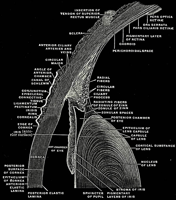
Lens Capsule
The lens is an elasticated capsule which assumes a non-spherical, globular shape when not being pulled into tension
by the action of the zonular fibres - a process connecting the lens capsule to the ciliary body. There are three
sets of ciliary muscles in the eye, the longitudinal, radial, and circular muscle - which work together to constantly
shape the lens to focus light on to the retina.
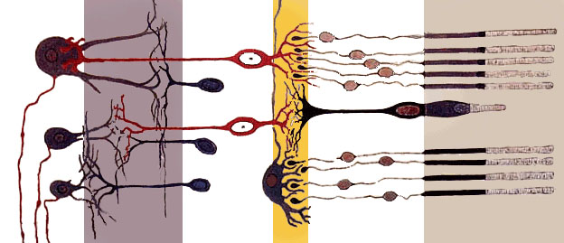
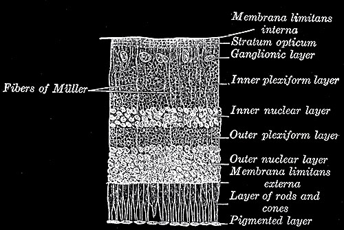
Retina The retina is constructed of several layers of neurons
in tissue on the inner surface of the eye. The retina, along with the optic nerve, are considered as integral parts
of the central nervous system (the brain). The retina can be considered much like a cmos device in a modern camera
or photographic film in an old-fashioned camera: light from the external world is focused onto it via the lens
to form an image.
.
Image from wiki - originally Gray's
Anatomy - creative commons licence
The retina is a complex, layered structure with
several sheets of neurons interconnected by synapses. Only the photoreceptor cells -rods and cones - are light-sensitive.
Electrical signals from the rods and cones undergo sophisticated processing by other neurons of the retina before
being relayed to retinal ganglion cells, whose axons form the optic nerve (see diagram left).
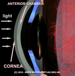
Cornea
Overview
Unlike the lens capsule, which is flexible and thus shape-changing, the cornea is a fixed curved transparent lens
covering the front of the eye (iris, pupil, and anterior chamber). It refracts light and accounts for about 66%
of the eye's focusing functionality; the remaining 34% is carried out by the flexible lens which acts to fine tune
the image onto the retina. As it needs to pass light unobstructed, it cannot possess blood vessels and must therefore
rely on nutrients diffused from tear fluid. It has uninsulated nerve endings which are extremely sensitive to touch.
Any contact to the cornea results in a reflex action to shut the eyelid and protect the eye.
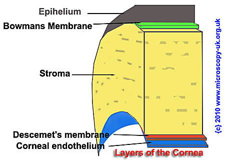
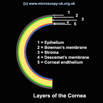
Layers
The cornea (in humans) consists of 5 layers:
The
Corneal Epithelium
This is a thin epithelial multicellular tissue layer of fast-growing cells. The cells are easily regenerated but
must be kept moist with tears. Dendritic cells (DC) and their immature counterparts, Langerhans cells (LC), are
highly specialized antigen-presenting cells (APC) located in the skin, mucosa, and lymphoid tissues. DC and LC
play a key role in the induction phase of contact allergenicity. Langerhans cells are present in The Corneal Epithelium
of the human eye.
Bowman's
layer This is a tough layer, 8 to 14 micrometers
in thickness, consisting of collagen fibres which protects the corneal stroma and help to strengthen and maintain
the cornea's curved shape.
Corneal
stroma
In addition to the known Langerhans cells in the corneal epithelium, at least three BM-derived cell subsets reside
in the normal corneal stroma. Dendritic cells (DCs) comprise a system of highly efficient antigen-presenting cells
(APCs) that initiate immune responses that protects the corneal stroma (*1-research
study paper).
The corneal stroma consists of about 200 layers of type I collagen fibrils with each layer being between 1.5 to
2.5 microns thick. Most of the corneal thickness is made up of this layer (90%). Theories of how the cornea maintains
transparency are complex and beyond the scope of this article, but in simple terms - two main theories exist. 1-
is that light scatter is cancelled out by a process called 'destructive interference' between light scattered by
individual fibrils of collagen in the stroma. 2 - The spacing between adjacent collagen fibrils in the stroma is
always less than 200nm, thus transparency is maintained. A detailed study into the transparency of the corneal
stroma has now been carried out and the results may be studied here at this off-site link provided by The National Center for Biotechnology Information
Descemet's
membrane
This is a thin cellular layer that serves as the modified basement membrane of the corneal endothelium, from which
the cells are derived (but in a different collagen structure). It is 5-10 microns thick.
Corneal
endothelium
The corneal endothelium is bathed by aqueous humour, not by blood or lymph like in many other body endothelia.
It look entirely different too. It is a simple single layer of mitochondria cells and regulates fluid and the transportation
of ions and small molecules between the membrane to the stromal area. The corneal epithelium cells do not regenerate
but stretch to compensate for dead cells.
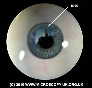
The
Iris
The iris functions much like the iris or diaphragm of a camera, controlling the amount of light entering the eye
by pulling on the pupil with muscle fibres (sphincter pupillae ) to contract it or by manipulating fibres (dilator
pupillae) to dilate (open) it. The iris really consists of two layers: the front pigmented stroma, with pigmented
epithelial cells beneath. The rear surface consists of a layer which is two cells thick containing pigment to block
light from passing through the iris into the retina of the eye. If this light was not obscured, the image at the
back of the eye would be overwhelmed with non-focused light. The iris is divided into two major regions: the pupillary
zone, which forms a bounday to the pupil, and the ciliary zone - the area forming the rest of the iris outto the
cilary body.
Ciliary
body The ciliary body is a thin vascular
(blood vessel-filled) middle layer of the eye situated between the sclera (the white of the eye) and the retina.
It is part of the uvea, which also includes the iris. The iris and ciliary body together are known as the anterior
uvea.
Uvea Part of the eye, consisting
collectively of the iris, the choroid of the eye, and the ciliary bod.y
Sclera
In addition to being continuous with the cornea, the sclera is also continuous with the dura mater, the outermost
of the three meninges, membranes covering the brain. The white of the eye connects with the dura mater at the optic
disc, located at the back of the eye. The optic disc is the location where the optic nerve exits the eye to carry
visual information to the brain. The sclera is perforated by many nerves and vessels passing through the posterior
scleral foramen, the hole that is formed by the optic nerve. It is Opaque to prevent light entering the eye except
through the pupil.
SUPPLEMENTARY RESOURCES
Human Eyes - position
A rotation of a 3d model showing human eyes in situ. In many ways, the organs of the human face/head - especially
the eyes and the internal top of the nose - should be considered as direct extensions of the CNS (central nervous
system). The eyes and nose are two areas of the human body which exhibit neurons composite to the brain, and which
are accessible to contact and study without invasive methods like surgery.
7Meg
bytes)of this rotation is available
for download here.
Eye Disease The eye is one of the most sophisticated
organs of the animal kingdom and a critical organ for survival. As with any complex mechanism, it is prone to many
issues even if slightly damaged or malformed. This article is more about the anatomy of a healthy human eye, and
therefore cannot really offer the very specialised information required to inform accurately about all the diseases
which can affect the eye. We therefore direct you to a list of human eye diseases on wiki
where you can explore all or any
of the topics you are interested in.
Additional Images
Above: image from wiki - originally Gray's Anatomy - creative commons licence
Credits Anatomy 3D model
data and .obj files from Anatomium Information derived from collating
and proving data from a variety of sources on the internet and from anatomy books offline. Where indicated, all image and video
content is copyright - mol smith - www.microscopy-uk.org.uk
Images which are not copyright are public domain or creative commons licence and are marked as such.
Comments or requests
for expansion of this article should be made to the author: mol smith
 .
.