by Mol Smith 2010  

Anatomy at Mic-UK
Human Heart


Return to Index


Content:   

 Location Parts Valves Timing Oddities    
    Disease Coronary Disease Cardiomyopathy Cardiovascular Valvular Disease    
    Animationa  HQ Movies Diagrams Links Credits    


Physical
Perhaps one of the greatest miracles of nature, is the creation of the human heart - a sophisticated pump designed to work without fault for approximately 90 odd years. In one lifetime, it will beat over 3.1 billion times and shift over 50 million gallons of blood in 80 years. If it misses just one beat, you will feel it as a momentary light palpation in the chest. It weighs approximately 325 grams (11.46 ounces - 0.72 lbs) in males and 275 grams in females.The heart pumps around 70 ml of blood per stroke (about 2.4 onces), and beats approximately 72 times a minute.

Location and Activity
The heart is located in the middle of the breast just beneath the breastbone which protects it. Slightly more of the heart projects out to the left hand side (as perceived by the heart owner). In many ways, it should be considered part of the lung/heart couplet as its location right next to the lungs is vital to its working.

The heart has two pumping circuits: one is pumping the blood out through the Aorta artery and into the main arteries and around the body; the other is pumping blood in a circuit through the lungs where blood is resupplied with Oxygen and returned to the heart to be transferred to the primary circuit. The heart provides the motion to force oxygen enriched blood through a massive network or arteries, blood vessels, capillaries and veins - thus supplying all organs and elements of our body with food and energy.

Construction
The heart has 4 main chambers called the right atrium, left atrium, right ventricle, and left ventricle. These are muscular bags that contract to
squeeze/push blood onwards. The left ventricle (chamber) has the thickest walls as it does more of the pumping. The chambers are arranged in pairs with the 2 atrium consisting of thin walls receiving blood from the veins. Each atrium pumps blood through a valve into its associated thicker-walled ventricle, which in turn - pumps the blood into a main artery. The sequence of operation is as follows:-

  • 1) oxygenated blood from the lungs arrive to the heart in the pulmonary veins

  • 2) It enters the Left Atrium

  • 3) The Atrium contracts and pushes the blood through the mitral valve into the left ventricle.

  • 4) The mitral valve closes and the left ventricle contracts pushing the blood out through the aortic valve into the aorta artery.

  • 5) Blood returns depleted of Oxygen from the body through a large vein into the right atrium.

  • 6) The right Atrium contracts, pushing the blood through tricuspid valve into the right ventricle.

  • 7) The right ventricle contracts - sending the blood out through the pulmonary valve into the pulmonary artery through the lungs where it is re-oxygenated.

  • 8) It returns to the heart (1) above and the cycle is repeated.


The 4 valves operate automatically opening and closing the valves to ensure pumping continues in one direction.

The Valves
The Pulmonary and Aortic valves are very similar with each having three leaf-like cusps made of tough, yet thin, fibrous tissue. The mitral and tricuspid valves are more complicated but similar in structure, with the mitral valve possessing two leaf-like cusps, and the tricuspid valve having three. These valves sit in in a ring with the free edges touching each other and able to close the passage between the ventricle and atrium. These edges are also attached to a set of fine strings (chordae tendinea) passing down into the ventricle. These prevent the valve from springing backwards when it is under pressure (like stays).

Timing
The timing of the valves opening and closing synched to the 4 chambers contracting and pumping is a critical process. With each heartbeat, both the atria contract together charging the ventricles with blood prior to the ventricles contracting together. Initial control is affected by the sinuatrial node in the right atrium. impulses from it pass through the muscles of both atria causing them to contract. At the junction of the atria and the ventricles, is another secondary node (the atrioventricular node) which delays the impulses before passing them on and down through a bundle of conducting fibres (called the Bundle of His) into the ventricles, causing them to contract after the two atria.


Oddities
After heart transplants, some receipients have reported changes in their behaviour such that they begin to favour activities or foods which the donor had favoured while alive. This has led to speculation that the heart may contain a degree of memory cells retaining traits from the donor. No scientific evidence has yet been found to support this theory. All cells do in fact possess some kind of latent memory but this is at a low level of functionality more to do with cell reproduction and immunity.


Diseases

Overview
Like all organs of the human body, the heart can suffer from a range of malfunctions and diseases. Since the heart is a highly critical organ, anything less than a perfect healthy heart can lead to serious consequences for the owner. Here is a non-exhaustive list of possible problem-causing conditions:

Aortic aneurysm
Coronary heart disease
Cardiomyopathy
Atherosclerosis
Congenital heart disease
Corpulmonale, a failure of the right side of the heart.
Hypertensive heart disease
Left ventricular hypertrophy
Valvular heart disease
Endocarditis
Myocarditis

Coronary Heart Disease
Coronary heart disease a term  that describes what happens when your heart's blood supply is blocked or interrupted by a build-up of fatty substances (atheromatous plaques) along the walls of coronary arteries that supply the myocardium. If your coronary arteries become narrow due to this build-up, your heart will start receiving a restricted supply of blood.  the blood supply to your heart will be restricted. This can cause angina (chest pains) or if they become completely blocked - a myocardial infarction, commonly called 'a heart attack'.

In the UK, 1 in 4 men and 1 in 6 women are dying through this disease, with over 300,000 people having a heart attack each year and over 100,000 dying in the UK, and approximately half a million in the united States.  Often, simple lifestyle changes, can make considerable improvements in the outcome of Coronary Heart Disease.

Cardiomyopathy
This is a disease of the heart muscle itself: the myocardium.  People with cardiomyopathy are often at risk of arrhythmia and/or sudden cardiac death. The normal functioning of the heart muscle may be affected by a variety of causes including genetic, alcohol and drug abuse, nutrition, inadequate oxygen delivery, diabetes, hyperthyroidism, excess accumulation of iron in other organs - especially, the liver, and diabetes.

 
Cardiovascular Disease
Vascular disease is mainly caused by hardening of the arteries(atherosclerosis) due to a thickening of the artery lining from fatty deposits or plaques (atheroma). Therefore Cardio-Vascular disease covers a range of conditions which mainly affects the heart. Normally, this is through restricted blood supply, as a result of narrowing arteries. Studies have revealed that in women, cardiovasular disease is more likely to be related to disease of the blood vessels, whilst in men - it tends to be disease of the heart muscles themselves. Known or associated causes of cardiovascular disease include diabetes mellitus, hypertension, hyperhomocysteinemia and hypercholesterolemia.

Cardiovascular Disease is commonly used as a collective term covering all heat disease as well as disease of the blood vessels.


The following will increase your risk:

  • Smoking
  • High blood pressure
  • High blood cholesterol
  • Physical inactivity
  • Being overweight or obese
  • Diabetes
  • A family history of heart disease
  • Ageing


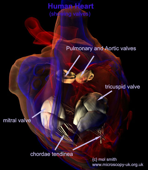
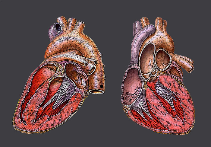
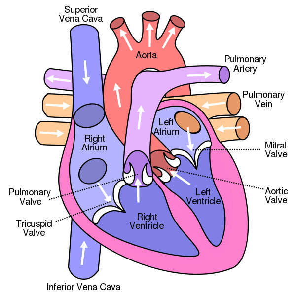
Valvular Heart Disease
There are 4 valves in the heart. As the term suggests, any disease of 1 or more of these valves will be termed as a Valvular Heart disorder or disease.

There are several types of valve disease:

Valvular stenosis
This occurs when a valve opening is smaller than normal due to stiff or fused leaflets. The narrowed opening may make the heart work very hard to pump blood through it. This can lead to heart failure and other symptoms. All four valves can be stenotic (hardened, restricting blood flow); the conditions are called tricuspid stenosis, pulmonic stenosis, mitral stenosis or aortic stenosis.

Valvular insufficiency.
Also called "leaky valve", occurs when a valve does not close tightly. When valves fail to seal completely, a quantity of blood will leak backwards making the heart work harder trying to compensate for the reduced push-through quantity of blood. Ultimately, it may not be able to compensate sufficiently and insufficient blood is circulated in the body.Depending on which valve is affected, the condition is called tricuspid regurgitation, pulmonary regurgitation, mitral regurgitation or aortic regurgitation


Heart Animations


Rotation of human heart showing valves


Rotation of human heart animated transparency


Multi-Rotation of human heart animated transparency

Higher Quality Animation Downloads
These may be used for personal use only. They must not be loaded onto other computers or networks without express permission from the author.

Please note : these animations were made using 3d Anatomy Data Models by
Anatomium

Rotation of human heart showing valves:  .mov file   download (576 kbytes)  .avi file  download (3.96 megabytes) 

Multi-Rotation of Heart .mov file  download (1.7 megabytes)      .avi file  download (11.6 megabytes) 

Rotation of Heart - Transparent    .mov file  download (1.3 megabytes)      .avi file  download (9.3 megabytes) 


Heart Diagrams


(above)
3D real heart - original image public domain. 3d Transform by mol smith
Transformation committed to the public domain.


Author: heikenwaelder@aon.at
Creative Commons Licence


Author: mol smith - image is copyright!


Diagram of Heart
Creative Commons Licence


Links
Wiki - Heart
British Heart Foundation
American Heart Association
BBC Heart Disease Site

Credits
Anatomy 3D model data and .obj files from Anatomium
Information derived from collating and proving data from a variety of sources on the internet and from anatomy books offline.
Where indicated, all image and video content is copyright - mol smith - www.microscopy-uk.org.uk
Images which are not copyright are public domain or creative commons licence and are marked as such.


Comments or requests for expansion of this article should be made to the author: mol smith


{c} Microscopy-uk.org.uk 2010 - all rights reserved. www.microscopy-uk.org.uk - e: help@microscopy-uk.org.uk