|
The Human
Lung
People tend to think of the lungs in a very simplistic
way, imagining them to be like a pair of balloons inflating and deflating. In fact, the lungs are far more sophisticated.
They are yet another marvel of Nature's sophisticated engineering and, along with the heart, form a critical major
organ of the body. Most Microscopists look at the lung in a series of sections through the lobes or Bronchi with
an optical microscope. Even at relatively low powers, it is fairly easy to determine diseased cells from healthy
ones.
Enemy
The greatest enemy of human lungs is cigarette smoke, not least because it contains carcinogenic substances which
increase the risk of cancers. Many people are also genetically susceptible to other compounds present in the smoke.
These compounds will cause a cessation of lung elasticity over time, resulting in ever decreasing lung capacity
and functionality, and ultimately causing a slow and miserable death. Although the lungs have mechanisms for clearing
dust and particles from their interior, it is not possible for them to unlock and clear carbon particles from the
cells. All damage caused by smoke is therefore irreversible! Any article about the human lungs would be less than
beneficial if it did not also cover some of the critical diseases, especially where they are preventable. So, I
will discuss both healthy and diseased lungs here. If you smoke (like me) you may prefer to negate the awful truth
about the consequences, but since I already suffer from COPD and my mother recently died of Lung Cancer, I can
only say to any reader that the sooner you give up the better. If you are so addicted (and I know what that is
like) consider switching to the new electronic
cigarette and you can continue with
your habit with a massively reduced risk of lung damage (almost zero risk).
Location
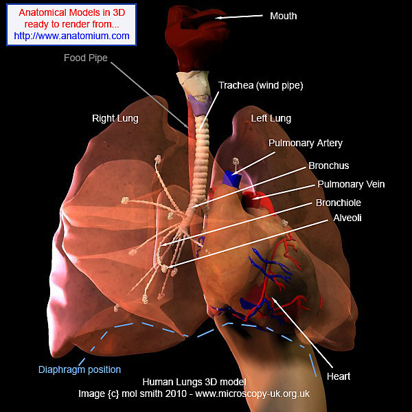
Almost the whole of your chest region is allocated as the home of your lungs and heart. You can see in my 3D model
below (if you have 3D red/cyan glasses, wear them now for true 3D), how the lungs and heart are prominently set
above the stomach and intestines.
The Lungs are in two halves with the Right side
(1st person perspective) larger than the left side - which (the latter) is smaller to make room for the heart next
to it. You can see the various major processes and the Lung's close association to the heart in the 3D model below,
where I have made the lungs semi-transparent to reveal the Bronchiole, Alveoli, and other processes inside.
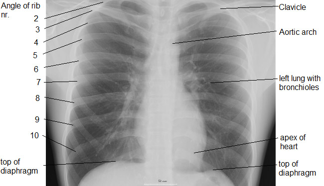
You may wish to consider the model against the x-ray below.
Processes
An outline of each of the main processes in the lung and also associated with both the lung and the heart are below.
Trachea
This is the basic windpipe with one opening arriving within the mouth and the other dividing off into the left
and right lungs forming the Bronchi.
Bronchus
/ Bronchi
The major two branches coming from the lower Trachea into the lungs (one into each lung). The right bronchus divides
into three Bronchi, which extend separately into the three lobes of the right lung. The left bronchus divides into
two bronchi which penetrate the two lobes of the left lung.
Pulmonary
Artery
The pulmonary arteries
are the only arteries in the mature human which carry de-oxygenated blood from the heart to the lungs. The blood
is reoxygenated and returned to the heart via the Pulmonary Vein, where it is transferred to the main blood circuit
carrying oxygenated blood to all parts of the body. See Heart Resources.
Pulmonary Vein
There are four pulmonary veins. They are large blood vessels which carry blood from the lungs to the left atrium
of the heart. There are two pulmonary veins from each lung. Unusually, these veins carry oxygenated blood - whereas
most of the other veins in the human body carry deoxygenated blood.
Bronchiole / Bronchi
(see Bronchus above)
Alveoli
The bronchioles (bronchi) divide repeatedly into many alveolar tubes lined with cuboidal epithelium. These terminate
in hollow lobed air sacs called Alveoli. There
are over 700 million alveoli present in human lungs, representing a surface area of approx. 85 square metres. The wall of each alveoli is just 0.0001 mm
thick, and on the outside is a dense network of blood capillaries which have originated from the pulmonary artery
and ultimately will rejoin with the pulmonary vein. Oxygen diffuses across the thin membrane represented by the
alveolar epithelium and capillary endothelium to pass into the blood plasma. It then combines with hemoglobin in
the red blood cells to form Oxyhemoglobin, whilst carbon dioxide diffuses ion the reverse direction into the alveolar
cavity. Note: the alveolar capillaries diameter is smaller than the diameter of the red corpuscles. This means
blood pressure squeezes the cell through the capillary, resulting in a slower progress and allowing more time for
the gas exchange to take place over a wider area of the blood cell.
Pleura
This is not shown on the 3D model. The pleura consist of two membranes separated for most of their area by a visceral
fluid (a bit like a super fine film) in contact with both surfaces, which allows the two membranes to slide smoothly
over each other. The two layers meet only at the hilum of the lung. (hilum : point at which the lung is connected to the trachea by its bronchus).
Any pain a person
may feel from their lungs, is probably originating from the Pleura as the lung interior does not contain pain receptors!
(Inflammation of the pleura or excessive fluid within the two membranes will cause pain! It is the surface tension
between the inner membrane and the outer membrane which hold the lungs open and prevent them from collapsing like
deflated balloons.
Diaphragm
This is not shown on the 3D model but is part represented by the broken blue line. The action of the diaphragm
is responsible for the movement of the lungs (inhalation and exhalation). The diaphragm is an area of muscle attached
to the chest cavity. When they pull on the cavity, the chest area expands (the ribs flex to allow this) and the
action of the pleural membranes (and the fluid sticking them together) pulls the lungs into expansion. When the
Diaphragm relaxes the chest wall contracts allowing the lungs to return to normal - exhalation / expiration occurs.
An interesting point to remember is that not all lungs are equal, in the fact that the lungs of birds, reptiles,
amphibians, and Invertebrates all differ from mammalian lungs (ours) and each other. More info here wiki.
The Optical Microscope can be used to recognise many diseases in sections of lungs of tissue. Before we move on
to look at this aspect through an optical microscopist's perspective, you may like to see an interesting 3D (red/cyan)
video of the lungs, and their position in the human frame by clicking on the thumbnail below.
Click thumbnail for 3 meg 3d video
The lungs are almost completely transparent so you can see their location
with respect to the heart and other organs more clearly.
Lung
Disease
As with any subject studied under an
optical microscope, what you see and determine correctly depends on several things. The first is understanding
what staining technique was used in the specimen, as different techniques can reveal different structure and processes.
The second, is often understanding what a specimen looks like when healthy and undamaged, and the third is knowing
something about interpreting flat 2 dimensional patterns and detail into a 3 dimensional comprehension. Is this
circular dot like that because of the way it was sliced? Is that curly thing with the thick blue perimeter really
a blood vessel, etc?
As we are now going to look at thin sections from the human lung, I think it is also a great opportunity to develop
these skills a bit further. So I will include some advice concerning interpreting staining results and how to recognise
structures in thin sections here. Don't think I am an expert at this, because I am not even a very good beginner:
I simply listened to the Brunel Microscopes Video and I have used their knowledge for what follows (used with kind
permission - see resources page).
The follow images were taken as stills, exported from a VHS video tape (a copy of a copy) so please excuse the
image quality. They are fit for purpose in this article though. To see a larger version of any example below, simply
click on the existing small image.
| |
|
|
|
|
| x 100 Healthy Lung |
|
Healthy Lung
- clear air sacs |
|
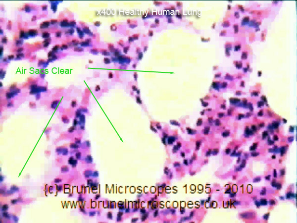
x400 Healthy Lung |
 |
|
These lung sections are stained with Haematoxylin wiki and
Eosin wiki which will stain cell nuclei blue and the cell Cytoplasm wiki pink. The healthy human lung, seen under a microscope,
will show typically clear air sacs (Aveoli wiki). These will be surrounded by cells of the lung comprising
small capillary blood vessels, bronchioles, and the Aveoli. The two slides (left x100) and (right x400) here can
help you understand this complex network, which is required for gas exchanges to take place. |
|
 |
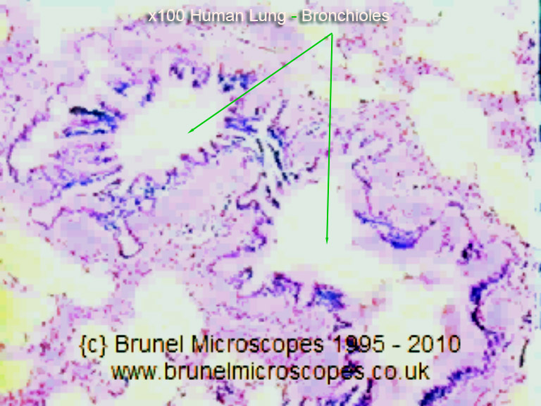
| x100 Healthy Bronchiole |
|
Healthy Lung
- clear Bronchioles |
|
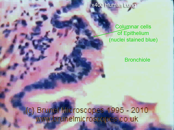
x400 Healthy Bronchiole |
 |
|
The Bronchioles (Bronchiola) can be seen here in cross-section
as large irregular empty spaces surrounded by the dark blue nuclei of Epithelium wiki cells which
represent the Bronchiole Wall. In the x400 slide (right) it is possible to see these cells are columnar in shape
with the nuclei at their base and the remainder of the cell cytoplasm extending away from it. Some of the cells
may appear less columnar, depending on their orientation, and where they were sliced in the thin section. |
|
 |
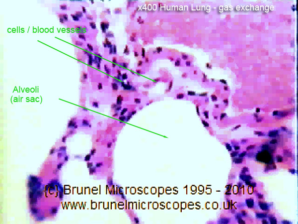
| x 100 Gas Exchange |
|
Healthy Lung - Gas
Exchange and cleaning |
|
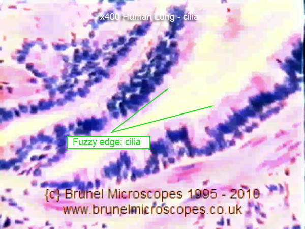
x 400 Cilia |
 |
|
 Gas exchange takes place in the Aveoli (tiny air sacs) through the network of fine blood
vessels (capillaries) surrounding each air sac. Oxygen passes into the capillary, and carbon dioxide along with
other waste gases are passed back into the air sac to be exhaled. In a healthy lung, the cells lining the Bronchioles
and air sacs are lined with fine cilia which 'waft' or sweep any mucus back out and upwards away from the lung
processes. They show up as fuzzy edges on the pink cell cytoplasm (see right) Gas exchange takes place in the Aveoli (tiny air sacs) through the network of fine blood
vessels (capillaries) surrounding each air sac. Oxygen passes into the capillary, and carbon dioxide along with
other waste gases are passed back into the air sac to be exhaled. In a healthy lung, the cells lining the Bronchioles
and air sacs are lined with fine cilia which 'waft' or sweep any mucus back out and upwards away from the lung
processes. They show up as fuzzy edges on the pink cell cytoplasm (see right) 
In diseased lungs, the cilia are destroyed, and mucus is cleared only by the action of coughing! |
|
 |
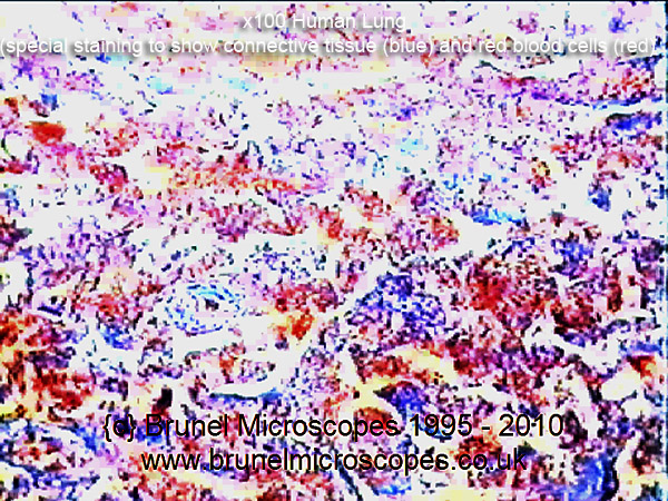
| x 100 Pneumonia Infection |
|
Diseased Lung -
Pneumonia |
|
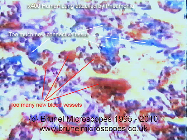
x 400 Pneumonia Infection |
 |
|
The specimens both left and right here have been stained
using a method called NSB Trichrome (off site), which shows
connective tissue as blue, and blood vessels and blood cells as red/orange. Here, too much connective tissue and
new blood vessels have formed as part of the body's (lung) defensive strategy to fight infection (get more white
blood cells in!). This severely interferes with gas exchange in the lung. The lung sections shown left and right
illustrate a lung infected with Pneumonia. Most changes to the lung are reversed once the infection has been medically
treated. |
|
 |
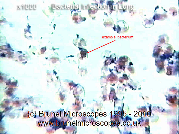
| x 1000 Bacterial Infection |
|
Diseased Lung - Bacterial
& TB Infection |
|
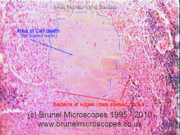
x 400 TB Infection |
 |
|
These slides are stained with Haematoxylin and Eosin.
Right slide shows area of Caseous Necrosis wiki
(cell death) where large areas of cells
lack the blue stained nuclei. This has been caused by an infection of Tuberculosis wiki bacteria
which can be seen at the edge of the damage. The bacteria causing TB are difficult to treat even with antibiotics.
The slide left shows
gram positive stained cocci -{spherical} bacteria infecting the lung. these are relatively easy to clear using
standard antibiotics.
See Staining Techniques
wiki regarding Gram Staining. |
|
 |
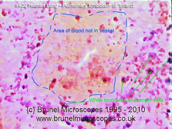
| x 400 Lung Infarction |
|
Diseased Lung
- Infarct and Miner's Lung |
|
x 100 Miner's Lung |
 |
|
The slide left shows large areas of lung tissue filled with red blood cells not contained
in blood vessels. They are surrounded by wandering scavenger cells and new connective tissue trying to repair the
damage. Where an area of lung loses its normal blood supply and starts to do die, the process is called an Infarct wk. In time,
the lung may repair itself by removing the dead cells. But not so in the slide of Miner's Lung (right)
where carbon (soot/coal) granules have filled lung structure and alveoli with deposits. The lung tries to clear
the damage with white blood cells, which fail and die in situ, increasing the area of non-functioning lung. Smoker's
lungs are also irreparably damaged this way. |
|
 |
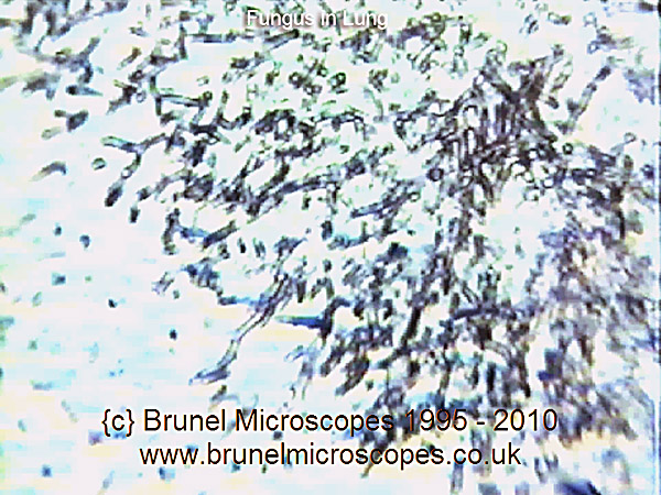
| x? Fungus in Lung |
|
Diseased Lung
- Fungal Infected Lung |
|
Movie of Lung Histology |
 |
|
Fungal infection in lung (typically Farmer's Lung wiki) caused
by spores from damp hay. Hyphae wiki can be seen as rosette clusters of stranded dark (Metallic Stained) filaments. These infections
are treatable.
If you click on the image to the right, you will be able to view a very low resolution version of a section of
film about the Lung extracted from Brunel
Microscopes Histology Video. |
|
 |
| |
|
|
|
|
|