|
Human Skin
A subject which can be studied easily
with a low powered optical microscope, is human skin. Although, most likely, the enthusiast microscopist, biology
or medical student will be using prepared stained sections - spotlighting interesting processes like sweat glands,
hair follicles, or sebaceous glands. We forget that the human skin is effectively the largest organ of the human
body. What an amazing job it does protecting all of our internal processes from the harshness of the external environment.
We are going to explore this extensive landscape here using 3D virtual models, along with images captured through
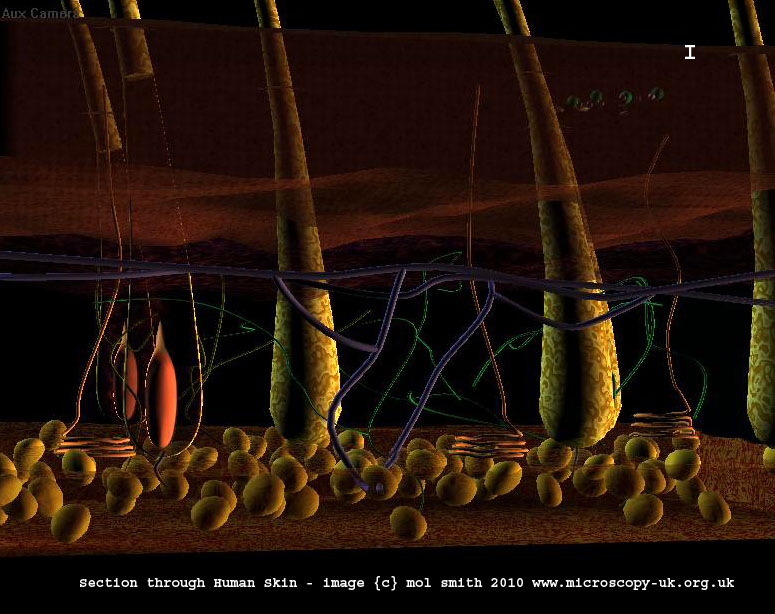
an optical light microscope. Below is a section taken through our 3d skin model.
First of all, I think this skin section model is not very accurate and only really serves to give a very artistic
impression, or interpretation, of the real processes in a section of human skin. For example, the hair roots below
the skin surface are not shown with their glands, which exist to lubricate the hair. I licensed it for around $50.00,
but to have obtained a very accurate 3D model would have set me back around $850.00. An accurate model exists here
at: 3D Science Frontier - but alas, I just don't have that kind of money :(
So here is the model...
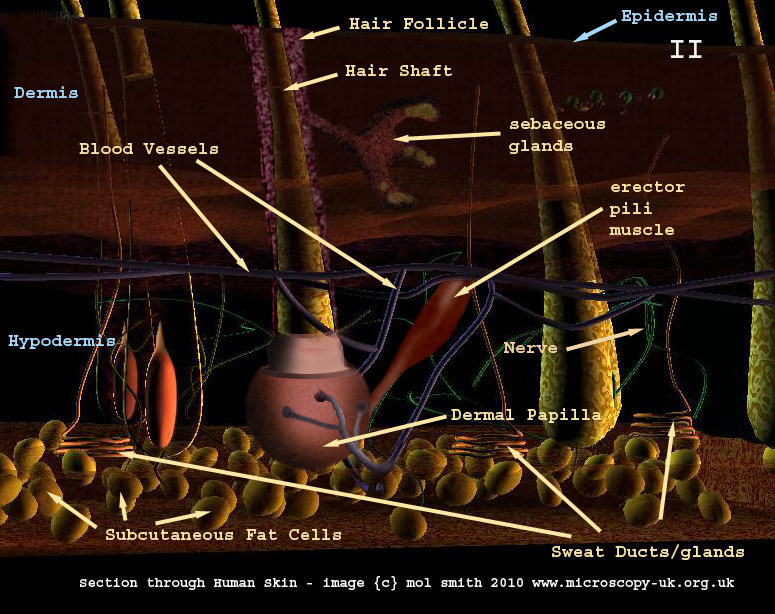
But this is not accurate. It requires a few additions,
so I have put them into the image by hand to show you the missing parts. I have briefly explained the function
of each labelled process below the image. It is interesting to note that when looking at a section through an optical
microscope, you may not see some of these processes because your thin slice was through an area where one or more
internal process was not in situ at that point. The advantage of a 3d model is that it can help you interpret a
particular slide section's processes.
This highly detailed sketch (below) of a skin section
is from an edition of Gray's Anatomy. I think it demonstrates all the processes in the human skin as accurately
as any modern (3D?) method. So, always remember, old skills of observation, patience, and attention to fine detail
can never really be surpassed.
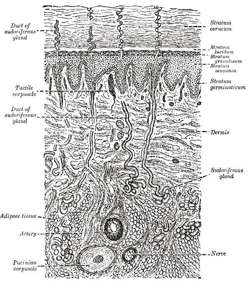
Grays sweat gland wiki
Expired copyright - public domain
Explanation
of Processes
| |
|
|
Epidermis
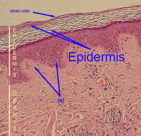
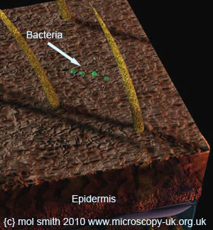
On the right, is an image
of a skin section taken through an optical microscope at about 10x magnification. The top layer consists of several
layers of dead skin cells. We are looking at the skin from the side. Orientation is assisted by the detail from
my 3D model (far right). The lowest level in the Epidermis consists of living mother cells which are constantly
dividing and moving up to the top, where they die, flatten, and transform into Keratin. This process takes approx.
26 days. The dead scaly cells are constantly shed. The surface of the Epidermis is covered with bacteria! |
 |
 |
| The Dermis and Epidermis are firmly joined together via a series of 'finger-like'
bulges in the dermis which 'plug' into sockets in the Epidermis -{A}. |
Dermis
The Dermis is comprised of collagen wiki
and elastin wiki fibres. Within this region are nerves, sweat glands, apocrine glands, sebaceous
glands, blood vessels, and the roots of tiny hairs. The nerves, and hairs, and sweat ducts penetrate the epidermis
but the blood vessels are confined to the Dermis only.
Hypodermis and Subcutaneous Fat Cells
The hypodermis is a layer of tissue that lies beneath the dermis and is mainly for fat storage. It consists of
loose connective tissue and lobules of fat along with larger blood vessels and nerves.
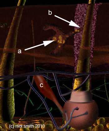
Sebaceous Gland
 |
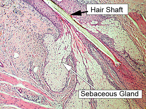
10x microscope image - creative commons
license - wiki
The sebaceous glands lubricate the hair shaft and skin. {a}- sebum is
an oily mixture of fatty acids, waxes, and steroids which pass into the hair follicle -{b}
and find their way up the hair shaft on to the skin surface. |
| |
|
Hair Follicle
and Hair Shaft
The hair develops from the papilla at the base of the hair follicle. This is nourished by tiny blood vessels. The
hair is composed of
cuboid epithelial cells which harden after impregnation with Keratin. The outer cortex of the hair will contain
various amounts of melanin - a pigment which determines hair colour.
Erector
Pilli Muscle
Attached to the follicle base is a tiny muscle - the erector pilli muscle - {c}.
Contraction of this muscle varies the angle of the hair to the plane of the skin surface. This serves two purposes:
control of the amount of warm air trapped between the hairs close to the skin surface (thermoregulation),
and increased perception of size of the human or animal in response to danger. The latter is due to all the hairs
standing on end which makes the organism look larger - a defence mechanism to help scare off would-be predators
or enemies. |
| Sweat Gland and
Duct |
|
|
There are two types of sweat glands, eccrine and apocrine.
The former are most common and are located in almost all regions of the body, whereas the latter are restricted
to public region, anus, arm pits, around the nipples, and on hands and feet. The glands are connected to sweat
pores in the skin surface - {ii} by way of coiled ducts {a}
and {i}. Blood capillaries transport water, urea wiki, and salts to the gland.
The apocrine glands release an odourless fluid which often turns quite smelly due to bacterial activity in the
fluid. Anti-perspirants and anti-deodorants contain zinc and aluminium compounds which destroy these bacteria.
The apocrine glands develop at puberty and their secretions occur during emotional or sexual arousal periods; the
fluids are far more viscous than that of the eccrine glands. |
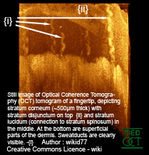
Still image of Optical Coherence Tomography wiki of finger tip. Rotation image below. Used via Wiki Creative Commons Licence.
|
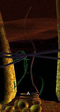
Section through skin model
showing sweat gland duct {A}. |
|
Dermal
Papilla
The term papilla is not a consistent term in scientific documentation and the naming of dermal papilla should
be used only for the connective tissue element, which is enclosed by the bulb of the hair follicle during the anagen
phase wiki, and which forms a compact ball of dermal cells underneath the "hair
germ" during telogen phase wiki, of the hair cycling and recycling process.
Nerves
The nerves terminate in both the dermis and epidermis
at specialised receptors (nerve endings) which transmit sensations of pain, heat/cold (Ruffini's corpuscle),
pressure (Pacinian corpuscle), and touch (Meissner's corpuscle).
Before we leave the subject of skin, and move on to the human lungs, I thought you might enjoy this little 3D video I made. You need those funny red/cyan
3d glasses to see this at its best.
|