![]() February
February
A nature walk for the microscopist presents more of a challenge at this time of year, with a few inches to a few feet of snow on the ground with little evidence of Spring yet. But if you look closely there's still plenty of colour and variety in the miniature world around us.
This month is hopefully a bit easier going after the introduction to the structure and identification of mosses last month!
Before you put your boots on, please read the important notes on collecting.
- nematodes (roundworms): these are tiny worms
which wriggle.
- rotifers: the commonest probably move in a leech-like manner
and were described in the January walk.
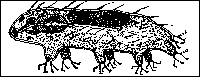
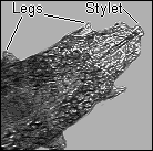
- water bears (tardigrades): these do in fact look like bears
with four pairs of legs and move in a lumbering fashion.
 To study under a high power microscope, transfer a
water bear to a microscope slide with a little water and cover
with a cover glass supported on a little vaseline to avoid
crushing. A water bear is illustrated on the left. They are
delightful creatures to study under a microscope. Water bears are
more accurately referred to as tardigrades, which are a class of
organisms containing about 350 species. They are usually 1mm or
less in size.
To study under a high power microscope, transfer a
water bear to a microscope slide with a little water and cover
with a cover glass supported on a little vaseline to avoid
crushing. A water bear is illustrated on the left. They are
delightful creatures to study under a microscope. Water bears are
more accurately referred to as tardigrades, which are a class of
organisms containing about 350 species. They are usually 1mm or
less in size.
They live in various habitats including damp moss, sand, freshwater and seawater. They have a well-developed head, and four fused body segments each bearing a pair of limbs which often have sharp claws. Most tardigrades eat plants, and feed by piercing plant cells with their stylets (spearlike structures near the mouth).
The most remarkable feature of tardigrades is their ability to withstand drying up and very low temperatures. Hence the reason why you can revive them from the driest piece of moss. When living conditions become unfavourable they go into a state of suspended animation. Specimens have been kept by scientists for over a year in liquid air (-190degC) and they have been successfully revived.
An unassuming but remarkable organism!
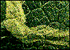
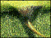 This thorn is on the midrib of
a bramble leaf (underside).
This thorn is on the midrib of
a bramble leaf (underside).
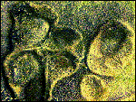
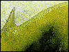
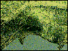 The leaf edge of a bramble
leaf.
The leaf edge of a bramble
leaf.
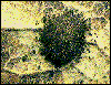
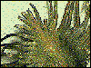 Dead seed and flower heads are
worth studying. This is a sepal-like bract from the seed head of
a knapweed (Centaurea).
Dead seed and flower heads are
worth studying. This is a sepal-like bract from the seed head of
a knapweed (Centaurea).
Go back to Walk Contents
Natural History Book Services A comprehensive on-line service with search facilities.
An excellent book with superb illustrations showing every
facet of microscopic life is:
'Microcosmos' by J. Burgess, M. Marten, R. Taylor.
Cambridge University Press, UK, 1987. ISBN 0 521 30433 4.
Tardigrades
An excellent introduction with further reading can be found in:
'An Introduction to the Study of Tardigrades' by P
Greaves. Microscopy, 1989, vol 36, p230-239. Microscopy is the
Journal of the Quekett Microscopical Club.
Tardigrade drawing from "Microscopic Freshwater
Life" by FJW Plaskitt, 1926. All other images by the author.
Fractal leaf created using Fractal Vision v2.5 by Dick Oliver,
Cedar Software.
Tardigrade image captured from the still frame of a VHS video.
Plant images were taken using a CCD camera attached to the
eyepiece tube of a stereo microscope using a x1 paired objective
with no eyepiece. Camera images were transferred to the PC using
a Creative Video Spigot capture card.
Image manipulation using Photostyler v2.0 software.
Comments to Dave Walker