 March
March
Seasonal snapshots of nature in close-up
Spring is a little way off in the north of England where the
author lives, but there are definite signs that the winter is
nearly over. The birds are singing and looking for nest material
and the catkins are out on the hazel trees. So why not join me
for a walk to see what can be found for study under the
microscope or hand lens?
Before you put your boots on, please read the important notes on collecting.
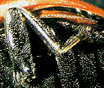
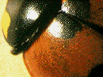 The
ladybird probably needs no introduction, as it is a common garden
beetle certainly in the UK. They are members of the beetle family
Coccinellidae and most species do a valuable job in
keeping destructive aphids and mites in check. But, have you ever
stopped to look at one under a 10X hand lens or low power stereo
microscope?
The
ladybird probably needs no introduction, as it is a common garden
beetle certainly in the UK. They are members of the beetle family
Coccinellidae and most species do a valuable job in
keeping destructive aphids and mites in check. But, have you ever
stopped to look at one under a 10X hand lens or low power stereo
microscope?
 Many species hibernate at
the same location each winter and often enter sheds or houses to
do so. In the recent milder weather they are becoming more active
and easier to find. All insects are fascinating when studied in
close-up. You don't need to be an entomologist just to admire the
wonderful texture and detail of the shell, legs, spines and
colour markings. The two images show a close-up of the head and
underbody of a seven-spot ladybird.
Many species hibernate at
the same location each winter and often enter sheds or houses to
do so. In the recent milder weather they are becoming more active
and easier to find. All insects are fascinating when studied in
close-up. You don't need to be an entomologist just to admire the
wonderful texture and detail of the shell, legs, spines and
colour markings. The two images show a close-up of the head and
underbody of a seven-spot ladybird.
There is no need to kill the ladybird to
study it. They often obligingly 'freeze' on the spot when touched
so can be viewed with ease. Flipping it on it's back will enable
a closer look at the legs and underbody. Return the ladybird to a
shrub outdoors when you have finished viewing it.
Go back to Walk Contents
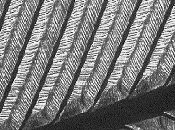 Occasionally you can find a pile of feathers and bones in the
countryside, where a bird of prey or fox has had a meal of a
bird. Feathers are always worth studying in close-up especially
if you can compare the small downy feathers and the larger
display feathers. There are a number of types of feather, which
have specialised functions ie insulation, flight display and
sensory reception.
Occasionally you can find a pile of feathers and bones in the
countryside, where a bird of prey or fox has had a meal of a
bird. Feathers are always worth studying in close-up especially
if you can compare the small downy feathers and the larger
display feathers. There are a number of types of feather, which
have specialised functions ie insulation, flight display and
sensory reception.
The typical feather consists of a central
shaft (rachis), with pairs of branches (barbs) forming a
flattened and usually curved surface called the vane. The barbs
possess further branches called the barbules, and adjacent
barbules are attached to one another by hooks which stiffen the
vane. In short, a marvel of nature's engineering!
Large feathers are easy to study under a
hand lens or low power stereo microscope. Try using transmitted
light, and also reflected light from various angles to fully
appreciate their structure.
Go back to Walk Contents
 A few trees start
to produce flowers towards the end of winter, the hazel (Corylus)
and alder (Alnus) are two of the earliest to flower. The
male flowers of the hazel (catkins) have been out in the north of
England for a few weeks, and are now bearing pollen. They are an
attractive sight and provide welcome colour to the countryside at
this time of year.
A few trees start
to produce flowers towards the end of winter, the hazel (Corylus)
and alder (Alnus) are two of the earliest to flower. The
male flowers of the hazel (catkins) have been out in the north of
England for a few weeks, and are now bearing pollen. They are an
attractive sight and provide welcome colour to the countryside at
this time of year.
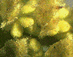
The catkins are worth studying under a low magnification, to
examine the structure of the flower. The anthers when ripe (shown
as yellow ovals in the image above ) will split to bear the
pollen which in turn is an excellent subject for examination at
higher powers under the microscope eg with a x10 to x40
objective. Just dab a little pollen with your fingertip on a
slide and cover with a cover-slip. You will have to vary the
focus to appreciate the structure because the depth of focus will
be small.
Pollen structure can vary widely between
species of flowering plant. We will investigate this in more
detail later in the year when more plants are in flower.
Why not join me next
month to study nature in close-up in Spring?
Go back to Walk Contents
Suggested reading
The large encyclopaedias such as Encyclopaedia
Britannica, are often a good introduction to flowers,
insects and birds. Most libraries should have plenty of books
that deal with these subjects in more detail.
Natural History Book Services
A comprehensive on-line service with search facilities.
Image details
Fractal cherry tree created using Fractal Vision v2.5 by Dick
Oliver, Cedar Software.
Images were taken using a CCD camera attached to the eyepiece
tube of a stereo microscope using a x1 paired objective with no
eyepiece. Camera images were transferred to the PC using a
Creative Video Spigot capture card.
Image manipulation using Photostyler v2.0 software.
Return to Walk Index
Comments to Dave Walker
© Onview.net Ltd, Microscopy-UK, and all contributors 1995 onwards. All rights
reserved. Main site is at www.microscopy-uk.org.uk with full mirror at www.microscopy-uk.net.