'WALK ON
THE WILDSIDE' SUPPLEMENT
Capturing images
of nature in close-up for display on the Web
by Dave Walker
As the October issue of the Nature Walk completes our first year of the Micscape magazine, we thought we would try to encourage you to take some of your own shots of nature in close-up. Why not have a try if you have access to the facilities and send us your favourite images. We would be delighted to compile a selection of them on Micscape.
If you are a student and your school or college has the facilities, why not start your own Web page of the nature in close-up in your area. We would be happy to provide a link to your site.
Briefly discussed below are a few ways of capturing 'close-up' images for the Web from magnifications as low as 2X to 200X and more, using as an example the simple equipment I use to illustrate the Nature Walks. It is very much a personal view based on my experiences and not necessarily the best way!
Four links to Micscape articles which discuss some aspects of close-up photography or video microscopy in more detail are also included.
Just take a
photo' ....
You may have a stills camera with macro facilities and may
already take close-up/macro photos of your local wild flowers,
insects or whatever interests you. Why not take two snaps of a
subject, keep one and if you would like your image on the Web
send us the other one (contact below). We can scan and upload it
and give you full credit for your efforts. Snapshot sized photos
eg 4x6 inch scan well usually. The secret is to get in close so
that the image fills the field of view. To get the depth of focus
put the camera on a tripod so you can use a slow shutter speed
and thus a high 'f' number - a flash gun may help. You could also
attach your camera to a microscope and take photos.
If you want to prepare images from colour prints for your own or school Web site but don't have a scanner, check the larger libraries or local college who may have scanning facilities. Note that if the images are for a Web page you don't really need to use an A4 flat-bed scanner. The flat-bed scanner can create a high resolution multi-megabyte A4 sized image .... but this will have to be reduced to a much smaller image a few tens of kilobytes in size for a Web page!
The author uses a hand-held (4 inch wide) 24-bit colour scanner which can easily scan the standard 4x6 inch colour snapshots. The images below were produced in this way using a resolution of 300dpi. Hand-held colour scanners are good value now.
Use your
camcorder (video camera / recorder)
If your family or school has a camcorder, many have 5-10X macro
facilities built in and this is an ideal way of capturing nature
in close-up outdoors or in an indoor setup with artificial
lighting. One of the advantages of the indoors setup is that the
electronic image can be sent straight to the computer and
electronically saved. You will require a video capture board for
your PC, which are quite reasonably priced nowadays. You can also
capture nature outdoors on the tape, and play the tape back home
while sending the video output to the PC to capture the
appropriate single frame.
If your camcorder does not magnify well enough you can get surprisingly good results with the additional aid of that humble 10X hand-lens we urge you to use. Read an article by Maurice Smith on how to do this, with examples of the results.
The video camera with or without macro facilities can also be used to photograph images seen down the eyepiece of a stereo microscope or higher powered compound microscope. You will have to carefully support the video camera above the eyepiece eg with a tripod and be careful not to damage the front lens element. Unlike older vacuum-tube based video cameras, solid-state CCD cameras are less susceptible to damage by high intensity light but it's still a good idea to slowly increase the microscope lamp intensity. You may need to experiment with the lens settings etc to minimise vignetting, the cut-off caused by the lens.
An article by Maurice Smith on using your camcorder for microscopy is in our Articles Library.
Using a
security type video camera
 If you were planning to do a
lot of image capture (or video microscopy) through a microscope
the security type video camera bought with a so-called 'C' mount
screw thread for the lens is a lot easier to use. This is the
sort of camera the author uses which was bought without a lens.
Unlike the camcorders they do not have tape recording facilities
but provide a video out signal which can be sent directly to a PC
with video capture board for saving stills or to a domestic
videotape recorder and TV/monitor.
If you were planning to do a
lot of image capture (or video microscopy) through a microscope
the security type video camera bought with a so-called 'C' mount
screw thread for the lens is a lot easier to use. This is the
sort of camera the author uses which was bought without a lens.
Unlike the camcorders they do not have tape recording facilities
but provide a video out signal which can be sent directly to a PC
with video capture board for saving stills or to a domestic
videotape recorder and TV/monitor.
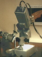
Admittedly the colour cameras are still quite expensive and are comparable in cost to a high quality camcorder but have a lot less versatility for general use. The author uses a Panasonic CL350 solid state (CCD) camera with 430 line resolution (horizontal). The camera is shown left attached with an adaptor (see below) to one eyepiece of a stereo microscope, using an external lamp for lighting.
Black and white video cameras are usually a lot cheaper and it is often possible to buy reconditioned ones from security firms, although some may have had a hard life so buyer beware! Note that older black and white cameras use 'Vidicon' or similar camera tubes rather than the lighter solid-state CCD chips found in camcorders. The former still give good results but are a lot heavier and can be damaged by overexposing to high intensity light.
If a security type video camera (or camcorder) is too expensive, I've noticed that solid-state miniature CCD cameras on a circuit board are now available from the larger electronic mail order catalogues for about half the price of a camcorder. These just require a low voltage power supply and some simple DIY to put a lightweight compact camera together for indoor fixed use. Most have a lens built in (not sure if they are easy to remove) which may or may not be beneficial for microscopy. In the UK Maplin catalogue for example, a high resolution colour camera costs 250 UK pounds. I've no idea how well these cameras work for microscopy, as they are designed for surveillance, so let me know how if you have experiences of using these cameras or thoughts on their suitability.
If you are considering buying a video camera either new or secondhand specifically for microscopy it is important to become familiar with the required technical specifications for microscopy work (see footnote).
An introductory article by Ron Neumeyer on using a security type camera for microscopy is in our Library.
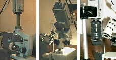
The author's
setup for capturing images
 The author uses a security
type camera without lens for image capture at both low and high
magnifications. Adaptors for this type of camera allow the camera
to sit directly on the eyepiece tube of both a compound
microscope and a stereo microscope. The simplest adaptor which
the author uses do not allow the use of the eyepiece, more
expensive adaptors do allow this. The image above left and right
opposite show the author's two microscopes setup in this way.
The author uses a security
type camera without lens for image capture at both low and high
magnifications. Adaptors for this type of camera allow the camera
to sit directly on the eyepiece tube of both a compound
microscope and a stereo microscope. The simplest adaptor which
the author uses do not allow the use of the eyepiece, more
expensive adaptors do allow this. The image above left and right
opposite show the author's two microscopes setup in this way.
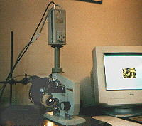
The video camera is attached to the compound microscope on the right with an adaptor on a vertical monocular tube. The video signal from the camera goes straight to the image capture board on the PC allowing the image to be displayed on the computer monitor. The composed and focussed image is saved on the PC for further image manipulation using widely available image processing software such as Photofinish or Photoshop. This software allows the image to be prepared for Web publishing.
Note that the sensor of security type video cameras is small (typically 6-8mm wide), therefore it is usually best to use this type of camera on the microscope without an eyepiece, or use a special projection eyepiece to allow a greater proportion of the field of view to be projected onto the image sensor.
The author possesses a very modest Russian Biolam compound microscope and Meiji stereo microscope with slide-in objectives. This type of stereo is quite cheap and are less than 150 pounds in the UK. The lamp for the compound microscope is an old enlarger lamp with an old camera iris in front, it gives a good even widefield illumination even at low magnifications.
If like the author, you have a 'moving limb' compound microscope (ie. the limb rather than the stage moves when you focus) the weight of the camera may change the focus of the image. However, the modern solid-state cameras are lightweight and this does not occur for the author's Biolam compound microscope, although it does occur for my stereo', so the focus is held using the focus knob while capturing a still frame on the PC.
If you are fortunate enough to have a trinocular head on the microscope you can keep the camera permanently on the microscope while still viewing through a binocular head. In the author's case I roughly compose the image visually down the stereo', then remove an eyepiece, attach the camera and do the final focussing and composition using the camera image displayed on the computer screen. For the compound microscope, the binocular head is removed and replaced with a vertical monocular tube. The magnifications achievable with both the author's stereo' and compound microscope are summarised in the table at the end of this article.
Using 35mm SLR camera
lenses etc with a video camera
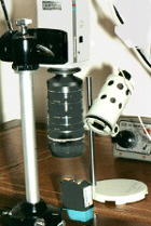 Low power macro shots using a
security type video camera is also possible without buying the
special 'C' mount lenses for the video camera. You can buy an
adaptor with an appropriate lens mount that allows standard 35mm
SLR camera lenses and ancillary macro equipment to be used. The
image left shows the setup through which many macro shots for the
Nature Walk page are taken. The lens is a Nikon 50mm standard
lens plus three standard extension tubes. The adaptor thus allows
you to use all your 35mm SLR macro equipment if you possess them.
Low power macro shots using a
security type video camera is also possible without buying the
special 'C' mount lenses for the video camera. You can buy an
adaptor with an appropriate lens mount that allows standard 35mm
SLR camera lenses and ancillary macro equipment to be used. The
image left shows the setup through which many macro shots for the
Nature Walk page are taken. The lens is a Nikon 50mm standard
lens plus three standard extension tubes. The adaptor thus allows
you to use all your 35mm SLR macro equipment if you possess them.
The video camera is mounted vertically on a normal camera tripod. This allows the coarse adjustment to be made using the focussing on the tripod pillar and using the 35mm camera lens for fine focussing .
An external high intensity light source will probably be needed for both incident light work with either a stereo microscope or the macro' setup above. A domestic high-intensity desk-lamp maybe suitable with either tungsten or quartz-halogen bulb. The latter will give better colour accuracy if this is important, although the author prefers the warmer tones of the tungsten lamps!
Magnifications possible
The magnifications possible with the author's setup is summarised
in the table below. Magnifications over a useful range of 1.6X to
425X on the monitor screen (6.4cm wide image) are possible, which
covers both macrophotography through to photomicrography.
| Set-up : Video camera (no lens) with ... |
Plus ... | Magnification on CCD chip (6.4 mm wide) |
Monitor screen mag. for 6.4 cm wide image |
| 50mm SLR camera lens |
'C' mount to 35mm lens
adaptor, plus 1-3 extension tubes |
0.16X - 1.3X | 1.6X - 13X |
| stereo microscope (no eyepiece) |
1X , 2X or 4X paired objectives | 1.8X - 5X | 18X - 50X |
| compound microscope (no eyepiece) |
3.5X, 9X, 20X or 40X objective | 4X - 43X | 41X - 425X |
Footnote:
Please note that the above is a general introduction to those
unfamiliar with the techniques of video microscopy and/or image
capture to produce images for 'Web' publication. There a number
of technical issues which I've glossed over but need to be
considered before investing in equipment. Seek expert advice if
necessary before any purchase.
Here are a few potential sources of advice.
1) Contact a regional microscopy society, who
may have members prepared to offer advice.
2) Posting a query in the sci.techniques.microscopy or
bionet.general newsgroup regarding cameras and video capture
cards should also produce a friendly and helpful response.
3) Try asking the biology department of your local college, they
may well do video microscopy and may offer impartial advice. They
are probably using professional equipment but may offer some
useful guidelines and contacts for second-hand equipment.
UK readers are particularly well-off in having a number of excellent microscopy societies who have members with a wealth of knowledge eg on video microscopy which is one aspect of image capture. A selection of articles from their publications is provided below.
So I hope I've encouraged you to try some close-up photography either on the macro' or micro' scale for the Web. Let me know how you get on if you do try and why not send us some of your results so we can display them, or even better start your own Web page and let us know the link!
Further Reading
1) Video microscopy using a TV camera fitted with a zoom lens
by D J M Thomson, Bulletin of Quekett Microscopical Club, 1991,
No. 17, pp. 12-13. This article describes the use of a simple
lens attachment in place of a compound microscope eyepiece to
project the full field of view into the camcorder and still allow
the use of the zoom lens of the camcorder.
2) Video microscopy using a TV camera fitted with a zoom lens: an improved system by D J M Thomson. Bulletin of the Quekett Microscopical Club, 1995, No. 25, pp. 9-10. An improvement on the original design which now requires no modification of the compound microscope optics.
3) Video-recording live protozoa by K Brocklehurst. Balsam Post (Postal Microscopical Society UK), 1995, No. 27, pp. 19-20. Describes trials on the use of a camcorder with a compound microscope.
4) An introduction to video recording at the microscope by A V Dodge, S V Dodge and K Jones. Microscopy, 1988, 36 (1), pp. 43-53. An excellent introduction to video microscopy for the amateur with particular emphasis on the basic information required by beginners. Discusses the choice of camera and particularly the use of ex-security black and white cameras. Lists UK suppliers. 'Microscopy', (now known as the Quekett Journal of Microscopy), is the Journal of the Quekett Microscopical Club.