I
wanderd lonely as a cloud
That
floats on high o'er vales and hills,
When
all at once I saw a crowd,
A
host, of golden daffodils;
Beside
the lake, beneath the trees,
Fluttering
and dancing in the breeze.
Daffodils
William
Wordsworth (1770-1850)
The crocus, hyacinth and daffodil are the most widely recognized
harbingers of spring in many parts of the world. In ancient times
the daffodil was considered sacred to the gods of the Underworld in
many Mediterranean cultures. Its scientific name Narcissus, derived from the Greek narke, means narcotic, and refers to the belief
at the time that the flowers scent produced a death-like sleep.
The flowers common name daffodil is thought to derive from a
corruption of the name of a different species, the white
asphodel. Affodyl became daffodil. Eventually the term
became associated with a large number of family members with the same
general structure.
Modern horticultural techniques have produced many new daffodils to
satisfy the appetite of the gardening public. Some have a strong
scent while others have practically none. All daffodils however,
share the same problem; all parts of the plant, (particularly the
bulb), are poisonous. The taste of the alkaloid containing sap is
so unpleasant however, that for most people, and foraging animals, a
single bite is more than enough! This sap is also deleterious to
other plants, and thus it is recommended that the cut flowers not be
mixed with other varieties in a vase.
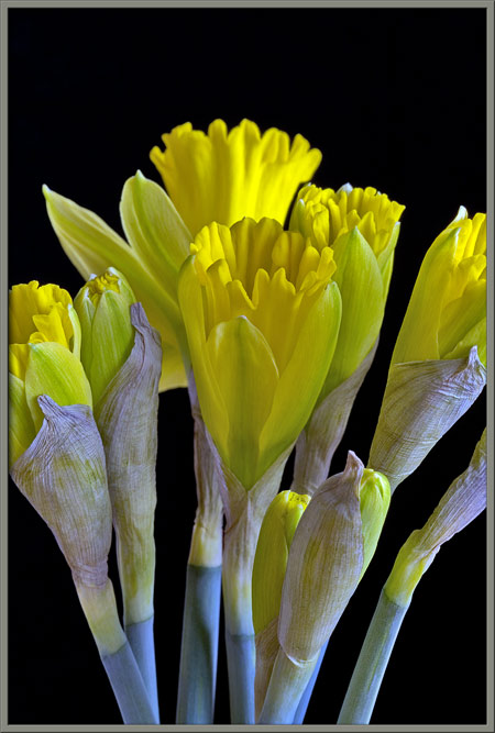
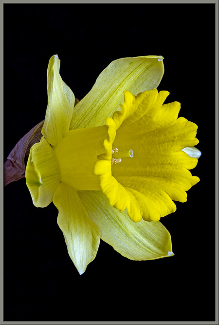
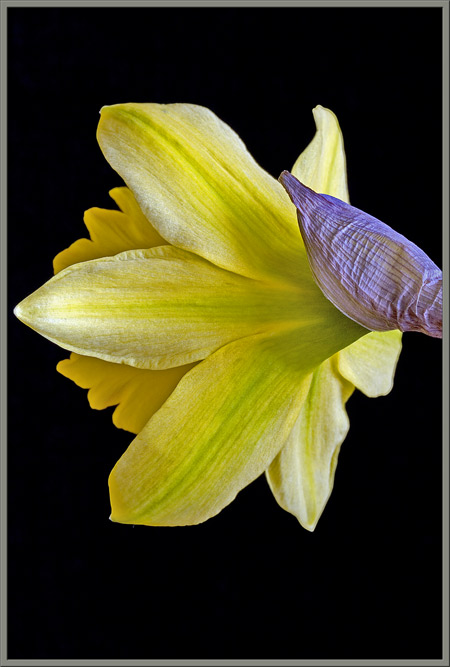
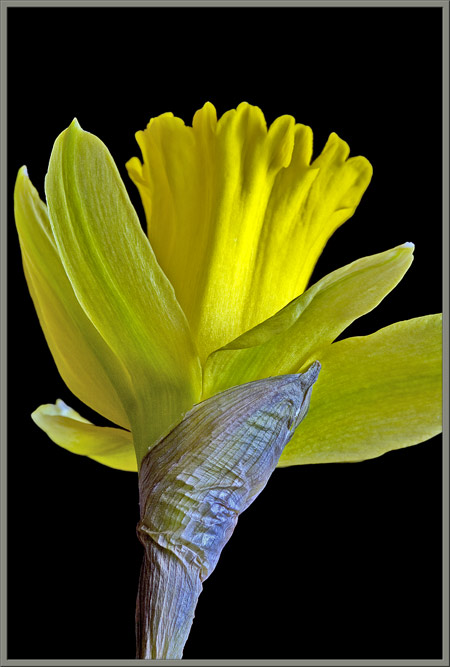
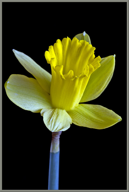
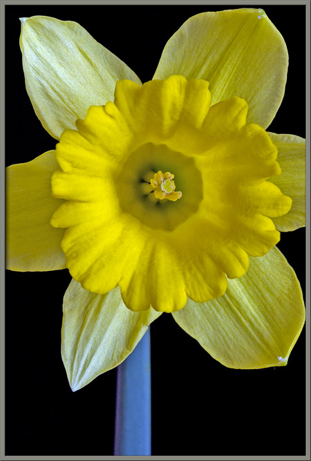
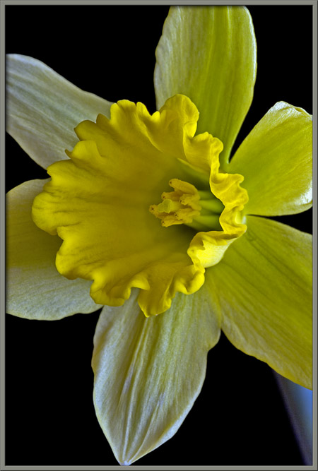
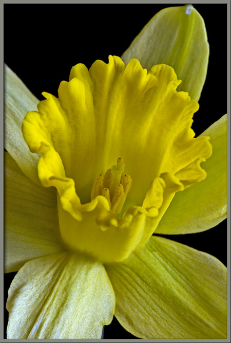
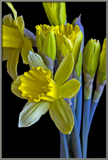
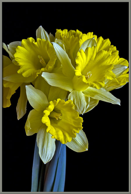
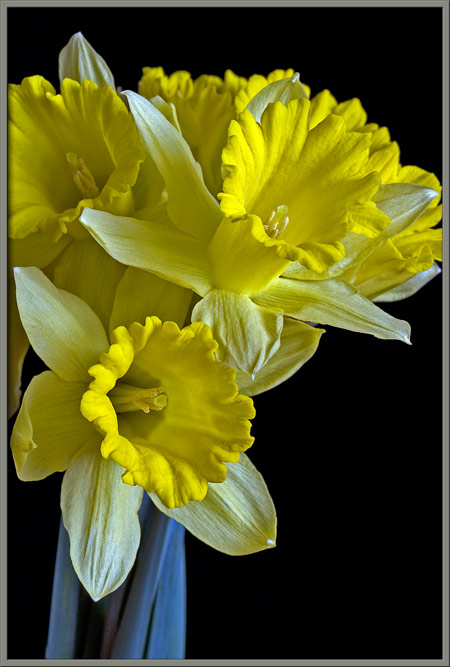
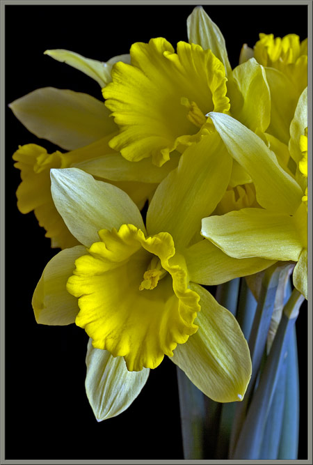
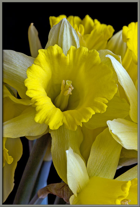
The four images that follow show the main parts of a daffodil
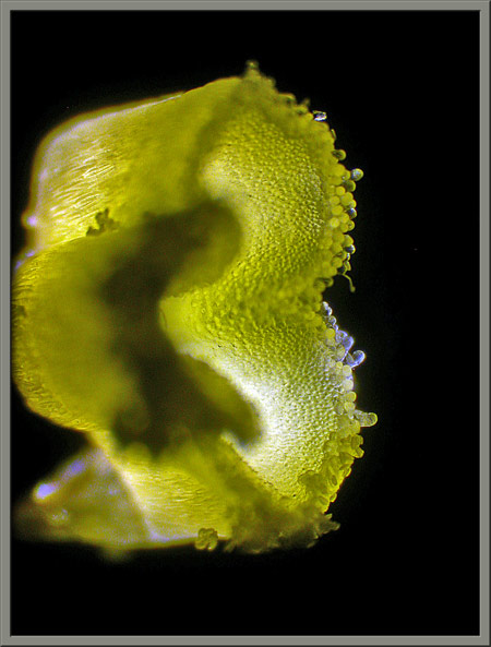
flower. There are six pointed petals that form the perianth. Extending outward
from the centre of the petals is the trumpet-shaped cup called the corona. In this instance, the
upper edge of the corona is rippled. The corona is much deeper
yellow, and is composed of much thicker tissue than the perianth.
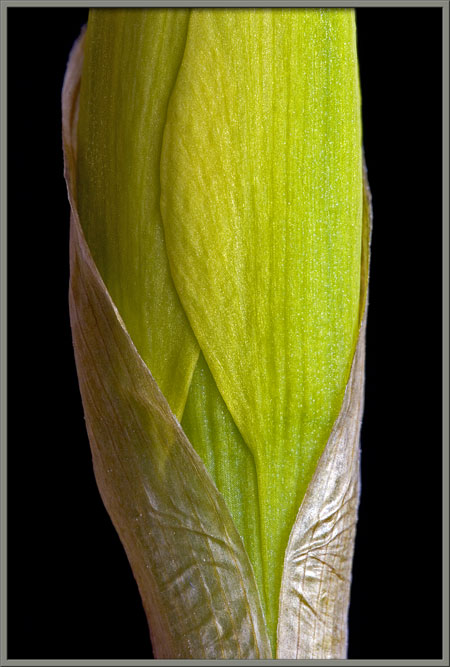
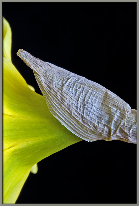
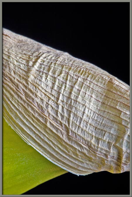
In the bud stage, the flower is protected by a thin, but strong,
membranous sheath called the spathe.
As can be seen below, this spathe splits lengthwise as the bud
increases in size.
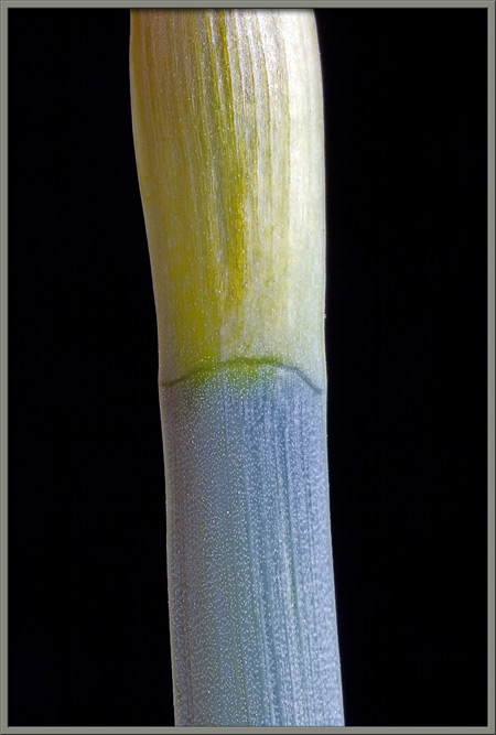
The base of the spathe is connected to the main stem at the dark green
ring seen below.
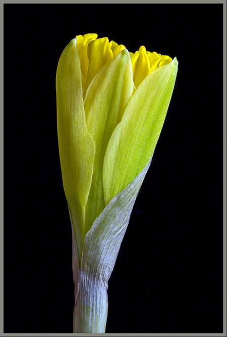
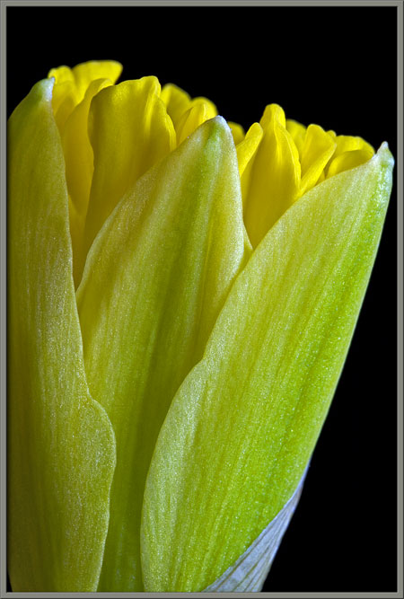
Two images follow that show the splitting, or unzippering of the
spathe as the opening bud swells.
Notice how tightly packed the petals are in the unopened bud.
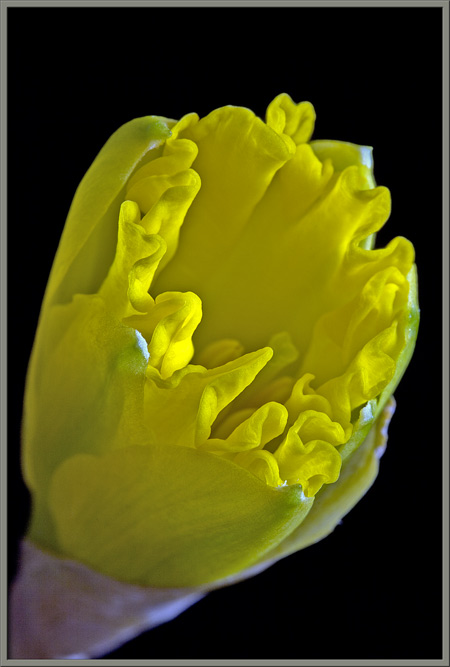
Eventually, the petals begin to separate, revealing the interior of the
cup portion of the flower. Notice the interesting yellow-green
shading in the underside of the petals shown in the left image.
A front and back view of a single flower can be seen below.
The spathe continues to enshroud the flowers base after blooming.
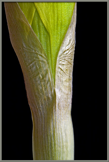
As you can see below, the spathe contains strong lengthwise ribs
combined with thinner, weaker tissue between the ribs. This
allows the rip that forms when the bud grows too large, to be a
straight one. The ribs prevent the rip from happening in a
crosswise direction.
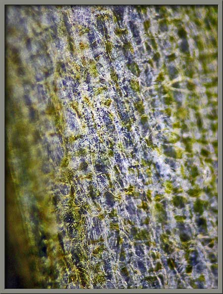
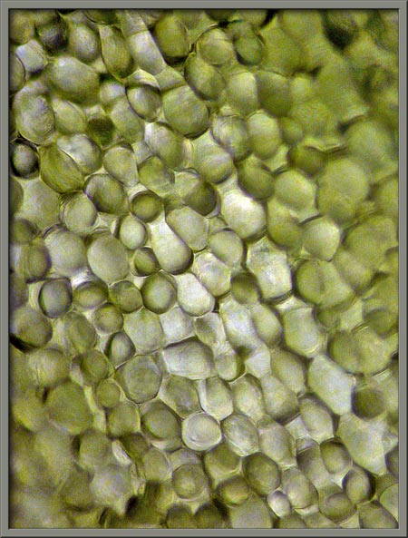
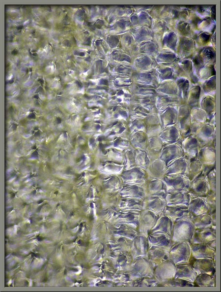
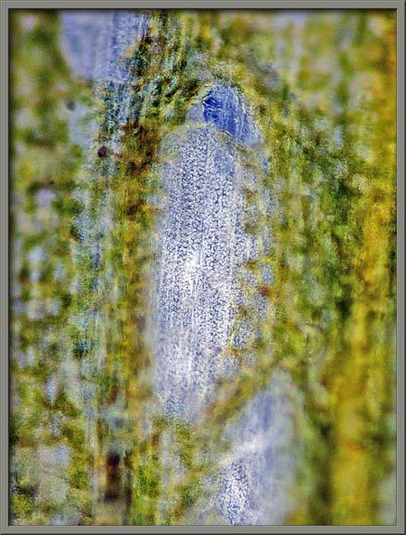
Under the microscope, the spathes cellular structure can be
observed. Note the fine hair-like filaments that cover the upper
surface in the image on the left.
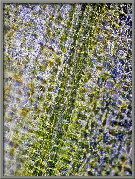
The cellular structure of a petal can be seen at right below.
This structure is not uniform over the petals surface. Some
areas are less yellow than others (left image), and other areas have
observable defects (right image).
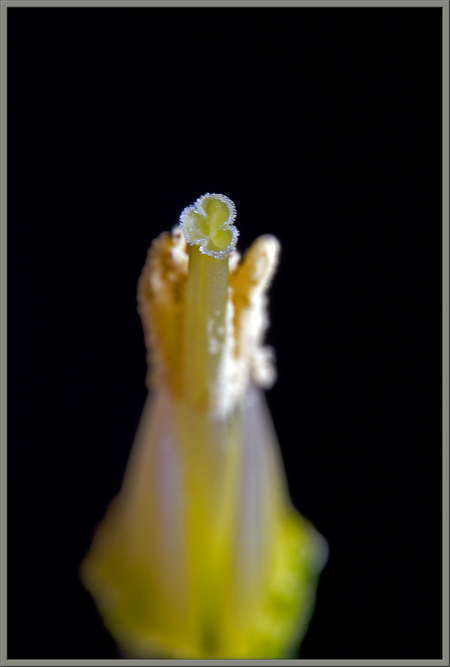
When a daffodil flower is viewed from the front, the reproductive
structures are clearly visible. At the very centre is the
light-green stigma (female
pollen accepting organ). Surrounding it are the tops of six
yellow anthers (male pollen
producing organs).
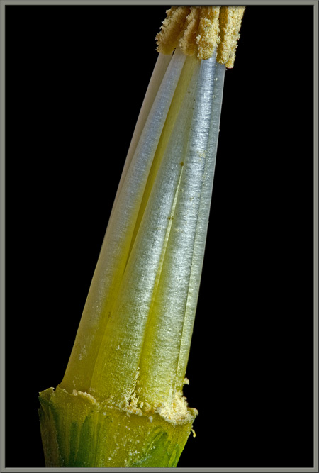
The angled view on the left below reveals the green filaments that support the
anthers. The pale yellow style
that supports the stigma is just visible in the right image.
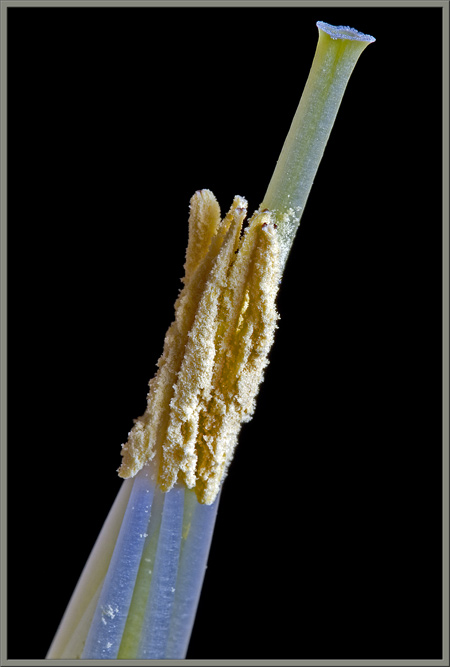
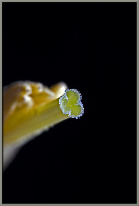
If the petals are removed from a flower, these reproductive structures
are easier to see. Starting from the top, the stigma connects to
the style, which is surrounded by the six pollen encrusted anthers,
which in turn are supported by their columnar filaments.
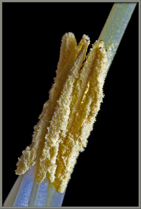
These columnar filaments are quite striking when viewed close-up.
The anthers seen in the image at right seem to retain the position
shown - close to one another and surrounding the style.
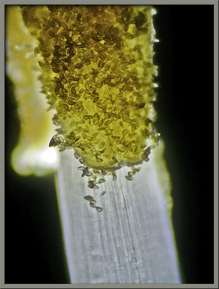
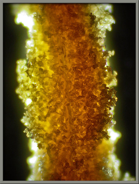
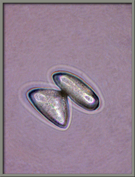
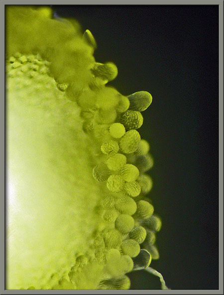
Under the microscope, the many tiny pollen grains that coat the surface
of an anther are visible. The anthers supporting filament can be
seen in the left image.
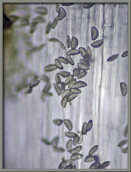
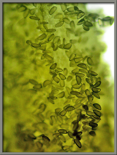
A higher magnification reveals the vaguely boomerang shape of each
grain.
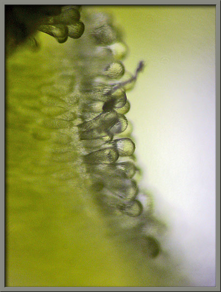
Phase-contrast illumination provides a different perspective.
Two views of the top of the daffodils stigma follow. The stigma
consists of three semi-circular lobes, each with a fringe of tiny
hair-like protuberances.
A view under the microscope of the top of the stigma can be seen below.
Much higher magnification reveals the nature of the hair-like
protuberances. From above they look like spheres, but in profile
they are indeed hair-like.
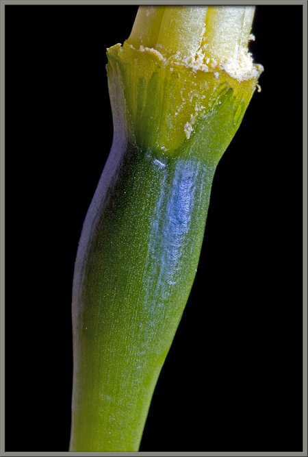
The style and filaments are connected to the top of the swelling in the
stem seen below. This is the daffodils ovary (seed producing
organ).
Daffodils belong to the Amaryllidaceae
family which contains about 1100 species. A large number of these
species are cultivated for their attractive flowers. The genus
Narcissus includes the daffodils, narcissi and jonquils.
Photographic Equipment
The macro-photographs were taken with an eight megapixel Canon 20D DSLR
equipped with a Canon EF 100 mm f 2.8 Macro lens which focuses to
1:1. A Canon 250D achromatic close-up lens was used to obtain
higher magnifications in several images.
The photomicrographs were taken with a Leitz SM-Pol microscope (using
dark ground and phase-contrast condensers), and the Coolpix 4500.