| COL's Renditioning of Blood A few images of blood
using COL illumination
By Paul James
(UK)
|
Editor's note: COL is circular
oblique illumination.
The author's four part series describing its creation
and use begins here.
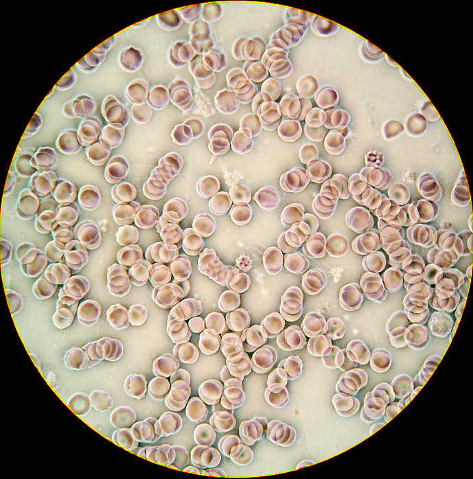 |
| A typical
full field view of blood cells in COL. The colour of both
the cells and background can vary depending on the objective
in use, it's na., and the width and 'thickness' of
the annulus.
|
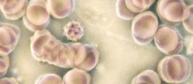 |
A crop of
the prime CCD image above ( 2560 x 1920 pixels = x 3000
on-screen ).
For screen size of 1024x768 pixels. |
 |
x 3000 (on-screen) Wild x 50 oil
Fluorite 1.0 na Objective
DF/COL using a
Heine condenser. Showing red corpuscles and
two
neutrophils which display their protoplasmic granularity.
The subtle
positioning of the Heine accounts for wide
variation of
effects of which this image is just one.
Note the
neutrophil's nuclei revealing typical 'Skull Eyes' look.
|
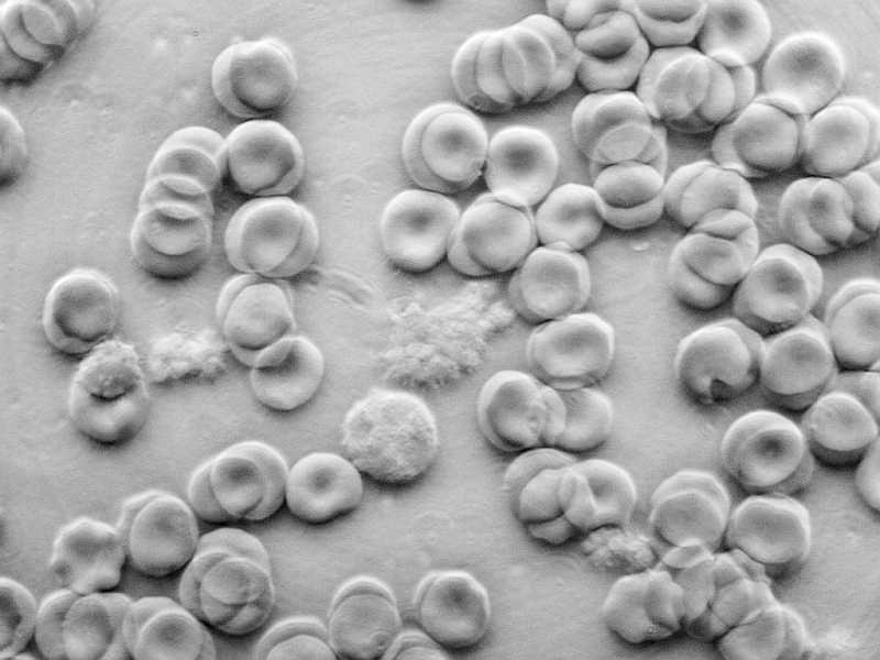 |
| x
2600 (on-screen) Wild x40 Fluotar 0.75 na This greyscaled image shows
a neutrophil and lymphocyte plus fragments of waxy looking
clotting material.
The cast shadow
'electron microscope look' has been brought about by
offsetting the
COL annulus, which often enhances contrast and helps
to define
imagery (and also
DUST) but does not suit all subject matter. This image
certainly shows what can be done
with a mere 0.75na
of objective aperture.
|
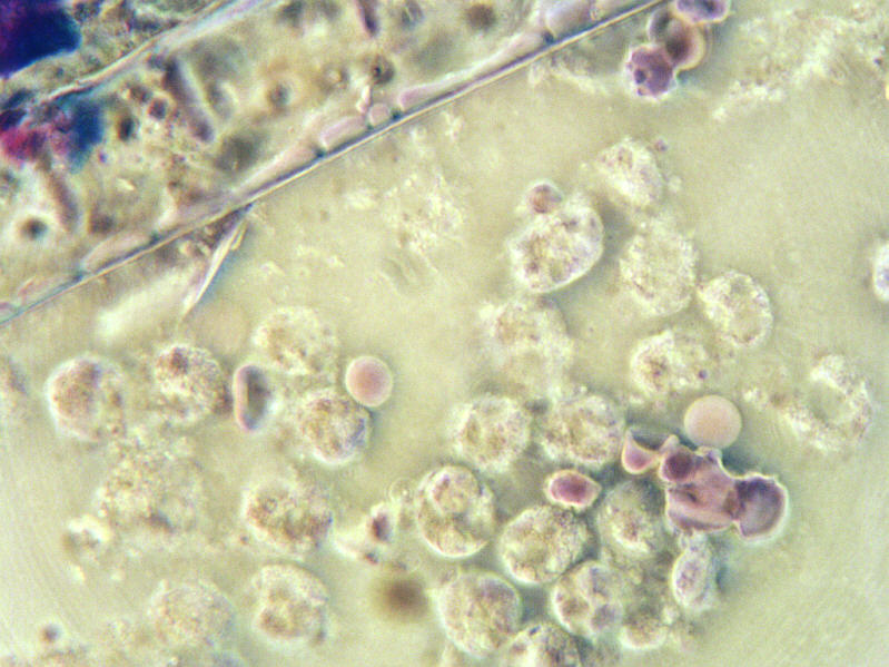 |
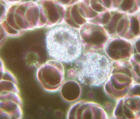
| x 1850
(on-screen) using Wild x 40 0.75 na Fluotar Objective in
COL. This is a sample of discharge from a wound. Many depleted neutrophils with
their characteristic nuclei in the field, and one or two
lymphocytes
and red corpuscles are
present too. A filament of 'micropore' dressing can be
seen near the top border.
|
NOTES
The blood smears
were simple untouched preparations below a coverslip which was
pressed firmly to reduce the film depth to allow COL to work at
its best. Magnification estimates were based on a simple
calculation using the average width of a red corpuscle which is
about 7.5 microns. Thus if the image of a typical corpuscle
measures 22 mm then when divided by 0.0075mm ( 7.5 microns ) the
amplification on-screen is approximately x 3000.
The images were
captured using a Minolta F300 digital camera above a Zeiss
Photomic stand. The camera's x 3 zoom and also the x 2 'Zeiss
Optovar' internal amplication unit was brought into use to
represent as large an image as possible on the CCD of the camera.
This effectively reduces the inherent noise of an image which
tends to be exacerbated by the lower light intensity of COL.
The magnification
in most cases is empty and excessive, but this was necessary to
illustrate the potential of COL using a difficult subject matter
without any preparation. As usual the live imaging was distinctly
superior to these images where the unique colouring effects of COL
and its inherently high contrast inducing properties excel.
The dreaded dust
spots in eyepieces are all too evident above and also
unfortunately the ringing problem with the Minolta lens, which is
exagerrated by COL........ an observation but no excuse!
The list of
specimens suitable for observation with COL is slowly
growing..........diatoms, butterfly scales, protozoans, bacteria
and blood films........enough to keep the observer busy for a
lifetime.
Microscopy
UK Front Page
Micscape
Magazine
Article
Library
© Microscopy UK or their contributors.
Published
in the May 2004 edition of Micscape.
Please report any Web problems or offer general comments
to the Micscape
Editor.
Micscape is the on-line monthly magazine of the Microscopy
UK web
site at Microscopy-UK