| An overview of the paintings of Christina
Brodie - Christina Brodie, UK |
Readers
of Micscape may be familiar with the ink and scraperboard drawings of
diatoms and radiolarians from my periodic illustrated articles.
These actually constitute one facet of my artistic output; for the last
seven years or so, my specialisation has been in botanical and natural
history illustration. Born in 1974, I originally graduated in
fashion and textiles, but am entirely self-taught as a botanical
illustrator.
Recently I completed work on an instruction book about botanical
painting, titled Drawing And
Painting Plants, to be published by A & C Black in October
2006. The book takes a novel approach in that not only painting
and drawing methods, but techniques such as dissection, collection of
plants, environmental studies, microscopic work and botanical
nomenclature are all covered. Students not only learn how to study and
paint flowering plants, but the whole plant kingdom, including
coniferous trees, fungi, ferns, mosses, seaweeds and lichens. This
teaching technique derives from adult education classes I taught for
several years at a local adult education centre in Somerset. For
Micscape's 10th Anniversary edition, I have elected to display some of
the paintings from the book for readers to enjoy.
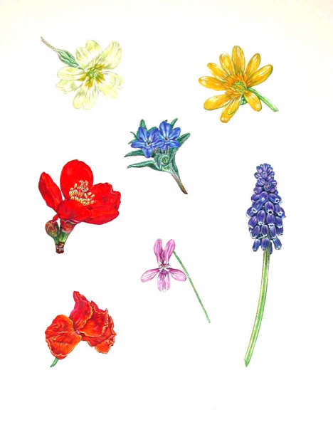
Coloured flowers: a
painting originally made to illustrate how specific colours of paint
can be matched to particular flowers.
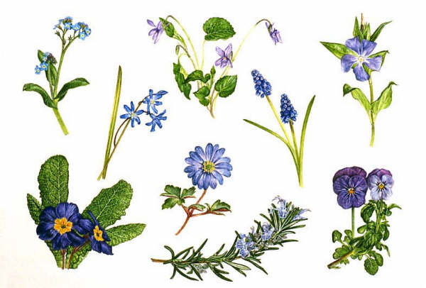
Blue
Spring Flowers: includes forget-me-not, Scilla, violet, grape hyacinth,
periwinkle, viola, rosemary, spring anemone, Primula species.
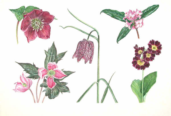
Red/Purple
Spring Flowers: includes hellebore, clematis, snakeshead fritillary,
Daphne, auricula.
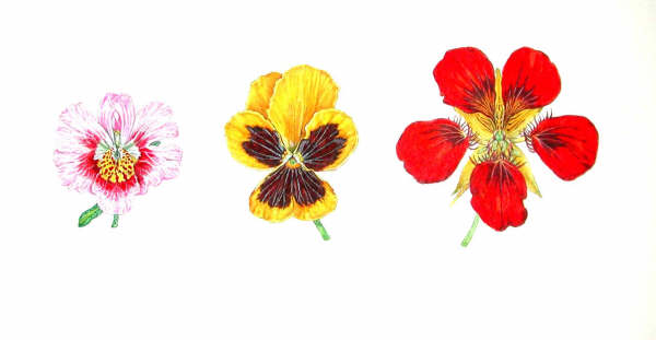
A painting made to show the
different patterns, or honey guides, on a flower's petals that lead
pollinators to the flower, in schizanthus, pansy and nasturtium.
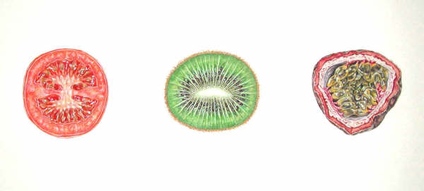
Sections
through fruit to show different ways in which the ovary is divided
(placentation): tomato, kiwi, passion fruit.
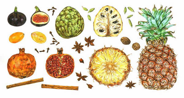
Tropical
fruits and spices: fig, cherimoya, pineapple, pomegranate, kumquat,
clove, cardamom, nutmeg, star anise, cinnamon.
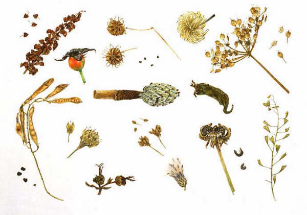
Seedheads, including those
of dock, clematis, umbellifers, crucifers, wintersweet, magnolia,
shepherd's purse, marigold, thistle, Hypericum, sweet pea.
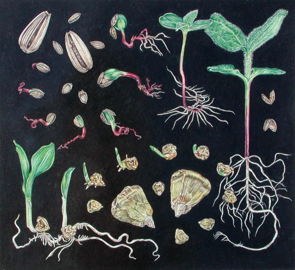
Development of monocotyledon
and dicotyledon seeds as shown in sunflower and maize.
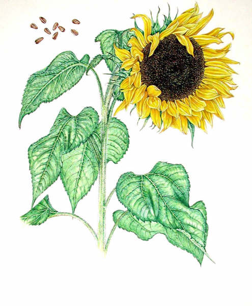
Sunflower.
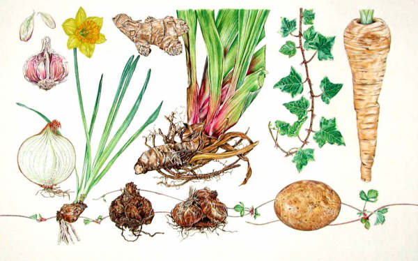
Different
types of root: gladiolus (corm), lily (scaly bulb), daffodil (tunicate
bulb), garlic (corm), ginger, iris (rhizome), ivy (adventitious root),
parsnip (taproot), potato (stem tuber), strawberry (stolon).
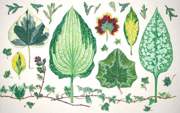
Variegated leaves: Euonymus,
ivy, hosta, zonal geranium, Aucuba, Pulmonaria, cyclamen and variegated
conifer Thujopsis.
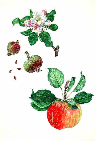
Study of apple "Discovery"
through flowering and fruiting stage.
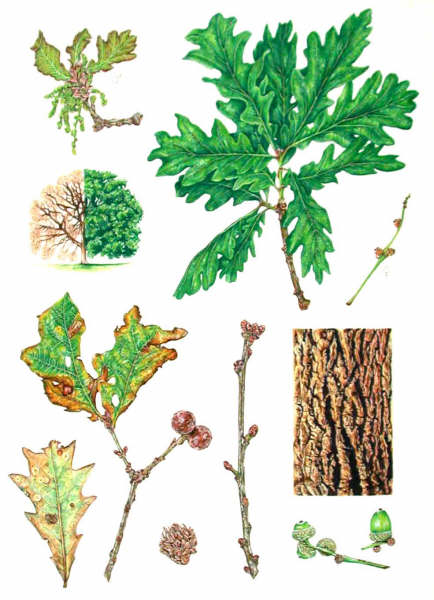
A comprehensive study of an
oak, showing male and female catkins, foliage, bark, acorns, winter
buds, autumn colour, the tree in summer and winter, and various galls.
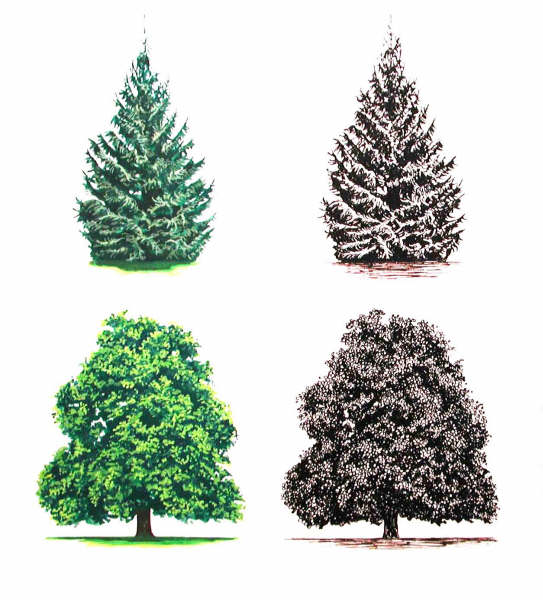
Rendering of tree shapes in
gouache and pen-and-ink.
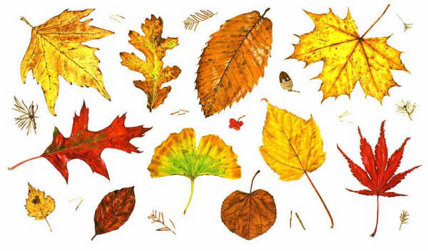
Autumn colour, as shown in
leaves from Westonbirt Arboretum. Illustrated are leaves of plane, 2
types of oak, 3 types of maple, sweet chestnut, katsura, beech, birch
and ginkgo, with fragments of larch, a spindleberry and an acorn.
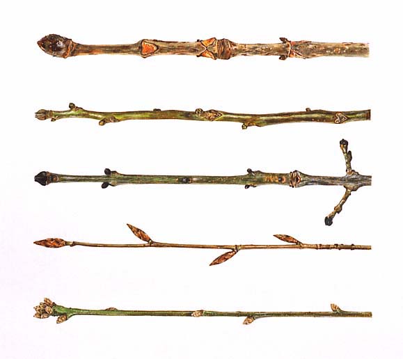
Winter Twigs 1 - Buds: horse
chestnut, walnut, ash, beech, oak.
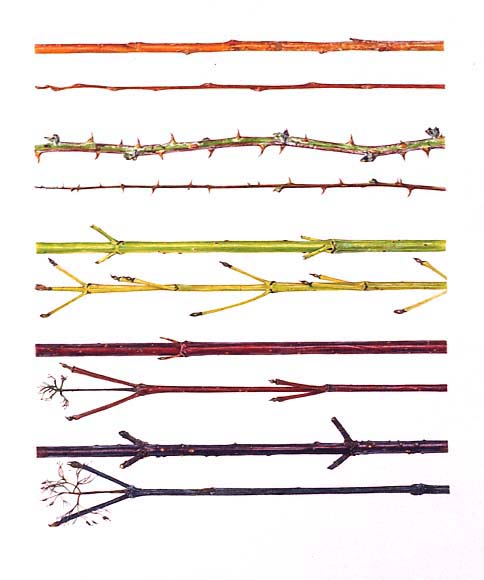
Winter Twigs 2 - Stems:
willow, bramble, 3 types of dogwood.
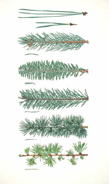
Needle-leaved conifers: 2-
and
3-needle pines, spruce, fir, Douglas fir, cedar, larch.
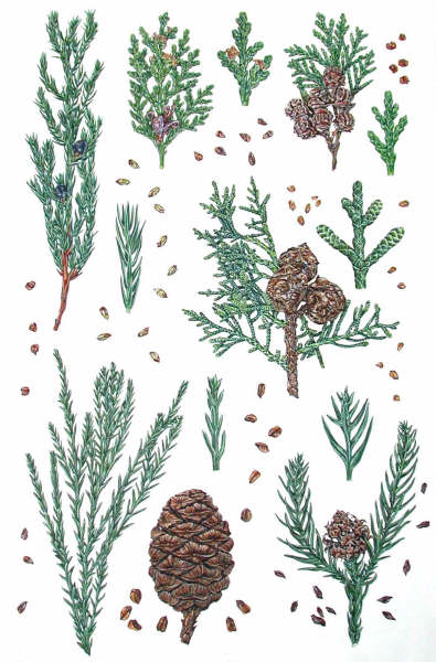
Scale-leaved conifers:
juniper, thuja, Lawson cypress, cypress, cryptomeria, sequoiadendron.
Illustrated with seeds and magnified shoot tips.
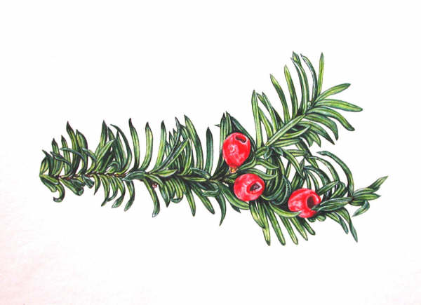
Yew, a conifer classed in
its own category.
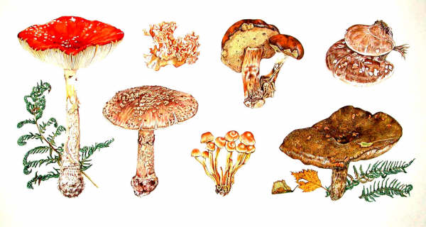
Fungi from a location on
Exmoor, including: fly agaric, blusher, cauliflower fungus, a bolete,
birch poplypore, Lactarius.
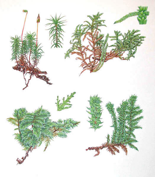
Different types of moss from
an Exmoor location, including a species of Sphagnum (upper RH corner),
and showing magnified views of the phyllids (specialized leaves).
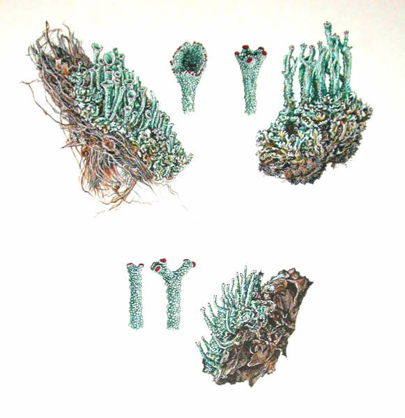
Cladonia lichens, the
magnified views showing branching and spore-bearing structures.
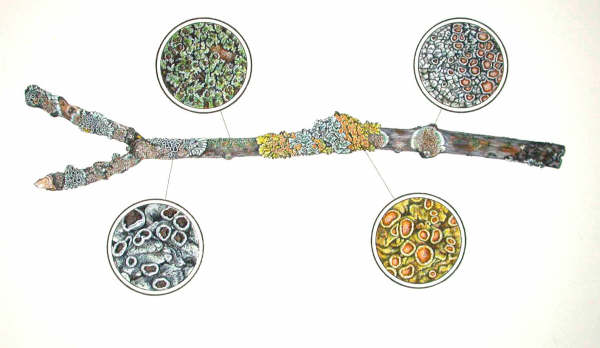
Crustose and foliose lichens
on an apple branch, with magnified views revealing the apothecia
(spore-bearing structures)
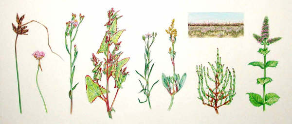
A painting showing how an
environmental study can be made of a rare floral profile from a
saltmarsh area and SSSI, using a combination of digital photographs and
sketches.
(*N.B.
Most of my work is from life and I generally encourage working from
actual specimens. However, I am in favour of the use of photography
where it makes practical sense, for example to record trees, bark, or
plants which should not be collected.)
The drawings seen below are
examples of the many (100+) ink drawings made to illustrate botanical
nomenclature throughout the book. They illustrate flowers that are
typical of particular plant families and that do not adhere to a very
simple form.
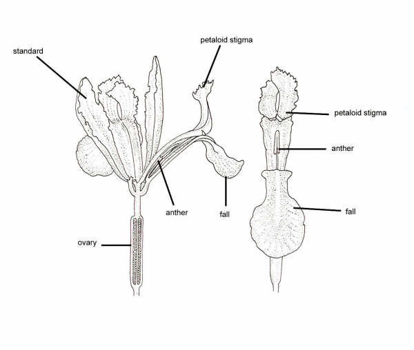
Iris half-flower and
one-third flower, showing the three-fold symmetry and how the stigma
and style are worked into the floral structure, deceptively appearing
to be a petal!
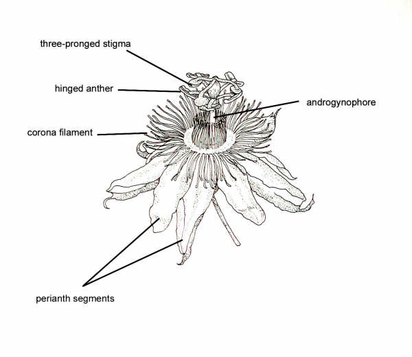
Passion flower. The male and
female reproductive structures are borne on a column called the
androgynophore, and surrounded by rows of corona filaments that form a
platform for pollinators.
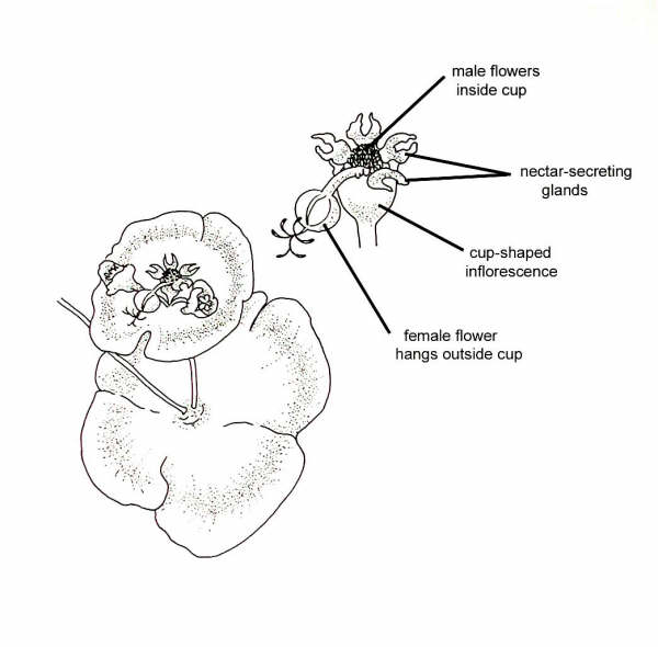
Euphorbia. The flowers arise
from the centre of a bract and are contained within a small cup,
surrounded by nectar-secreting glands. The male flowers are positioned
inside the cup, whilst the female flower protrudes outside.
This article gives a
snapshot of the book's content; in addition to the paintings (approx.
80), it will also feature numerous ink drawings (100+) illustrating
botanical nomenclature, and photographs (100+) to show painting and
scientific techniques.
See my website for contact details, at www.queen-christina.com, which also shows some of my
natural history illustration and design work.
November 2005
Images © Christina Brodie 2005. All rights reserved.
Microscopy UK Front Page
Micscape Magazine
Article Library
© Christina Brodie 2005.
Published in the November 2005 edition of Micscape Magazine.
Please report any Web problems or offer general comments to the Micscape Editor.
Micscape is the
on-line monthly magazine of the Microscopy UK web
site at Microscopy-UK.
© Onview.net Ltd, Microscopy-UK, and all contributors 1995 onwards. All rights reserved. Main site is at www.microscopy-uk.org.uk with full mirror at www.microscopy-uk.net.