DIATOMS IN THE DUNNY
(Tabellaria in the Toot)
(Lacrimaria in the Loo or Lav)
(Blepharisma in the Bog)
(Caloneis in the Can)
By Mike Dingley, Australia
A most interesting place to visit in order to examine
freshwater is inside your own lavatory. Remove the lid from the cistern
and peer inside. Around the water level will be a brown ring of 'gunge'
adhering to the cistern wall. Flush the toilet and this drains the water
from the cistern and leaves the brown ring easily accessible. Using a scalpel,
section lifter (see there really is a use for this) or some other flat
instrument, scrape a tiny amount of this 'gunge' and place on to the centre
of a clean microscope slide. Transfer a drop of water to this and if the
'gunge' is not too thick cover with a cover glass. If it is too thick tease
the fibres etc apart before placing the cover glass on top.
Lets go and have a look!
In the first example that I examined from my home I was
surprised to see large numbers of living organisms that had taken up rent-free
lodgings. I also thought about the cleanliness of our water supply but
didn't dwell on it for too long. The majority of specimens were diatoms
of various types. Now I'm not too cluey on diatoms but I was able to name
a few. Most were centric diatoms which when seen side-on gave the appearance
of thick tablets one on top of the other forming chains 3-8 cells long.
Other diatoms included Navicula sp., Caloneis sp., Tabellaria sp., Cymbella
sp., Rhoicosphania sp., Melosira sp. and Gomphonema sp. There were others
too small for me to attempt an identification.
There were many small ciliates swimming around making
it hard to keep following them and keeping them in focus (I must try and
use a smaller drop of water). Something that allowed ample time for observation
because they were stationary were dark orange circles or discs (great description
eh?). No they were not shells from Arcella but I am unable to identify
them. I wondered if they were cysts of some sort or another. I saw a rotifer,
Anuraea sp., as well as several amoebae of what appeared to be of the same
species. A few Oscillatoria, (blue-green algae or Cyanobacteria), and I
also saw the colonial desmid alga, Desmidium sp., which was 18 cells long.
There were some very active but un-identified nematodes present.
Swimming around quite nonchalantly were a few colonies
of Gonium sp., a colonial alga where each cell has a conspicuous flagellum.
These look quite nice when viewed using dark ground illumination. Another
colonial alga seen was the non-motile alga Dictyosphaerium which are loose
colonies of green cells in clear mucilage. A most interesting ciliate observed
was a large specimen similar to Paramecium which swam past and through
the field of view searching for food. The slow moving rotifer, Philodina
was moving in its leach-like or caterpillar action. Another fascinating
creature was busy walking over, under and on top of the fibrous matter.
It seemed to be searching for food and was similar in shape to the shell
noodles that we have sometimes for dinner. They had cirri which appeared
like they were walking on legs. It was either Euplotes or Aspidisca but
I will have to try and identify them at a later time.
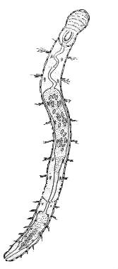
 A huge and interesting creature which came across my field of view was
an annelid worm being an Oligochaete of the family Aeolosomatidae. It measured
about 1mm in length, had a mouth in its head region (a good place for it
to have one!), and inside a cavity full of cilia which passed food particles
into its digestive tract. I observed food passing through its transparent
body and being ejected at the other end. Although the animal was transparent
it was nevertheless easy to observe as just under its skin were many bright
orange oil-like droplets, the purpose of which was unclear to me. It also
had spines radiating out from its skin along its length. I could also see
four diatoms within its intestine.
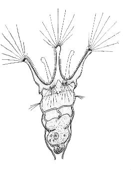
 The most beautiful creature that I came across and which has given me a
hard time identifying was a rotifer called Colotheca. It was trumpet shaped
and held to the substrate by a tapered foot which was, alas, hidden behind
some 'gunge'. It did not have a tube along its body but at its widest point
(the head) it had stunningly beautiful thin glass-like spines. These were
bundled onto four projections. They did not seem to do anything except
make the creature look beautiful, almost majestic. A tiny ciliate swam
into the funnel-shaped end and appeared to hit a barrier about of the way
inside the animal whereupon the animal closed its funnel-like rim like
a fist to prevent the ciliates escape. As quick as lightning the ciliate
disappeared in to the lower of the animal and was lost into the rotifers
more opaque looking body. Once this happened, the fist unfolded and its
long glassy spines gradually and gracefully protruded out into the watery
environment.
I have wondered whether you would get the same types of
animals and plants inside toilet cisterns from other areas of the country
or from older houses with old, antiquated pipes. It might be a good project
for someone to do. I have collected a few samples from motels that I have
stayed in but have had to preserve the contents of my collection and have
not had time to look at them. I also feel that it would be better to examine
samples when the contents are still alive. I did visit a motel once in
Victoria (Australia) where I could not remove the lid of the cistern. It
was a white plastic one but upon prising, twisting and banging I could
not remove the it so had to leave this area for another visit. So, have
a look in your cistern, go on don't be shy and you will be surprised.
Mike
Dingley
© Microscopy UK or their contributors.
Please report any Web problems or offer general comments
to the Micscape
Editor,
via the contact on current Micscape Index.
Micscape is the on-line monthly magazine of the Microscopy
UK web
site at Microscopy-UK
WIDTH=1
© Onview.net Ltd, Microscopy-UK, and all contributors 1995 onwards. All rights
reserved. Main site is at www.microscopy-uk.org.uk with full mirror at www.microscopy-uk.net.
 A huge and interesting creature which came across my field of view was
an annelid worm being an Oligochaete of the family Aeolosomatidae. It measured
about 1mm in length, had a mouth in its head region (a good place for it
to have one!), and inside a cavity full of cilia which passed food particles
into its digestive tract. I observed food passing through its transparent
body and being ejected at the other end. Although the animal was transparent
it was nevertheless easy to observe as just under its skin were many bright
orange oil-like droplets, the purpose of which was unclear to me. It also
had spines radiating out from its skin along its length. I could also see
four diatoms within its intestine.
 The most beautiful creature that I came across and which has given me a
hard time identifying was a rotifer called Colotheca. It was trumpet shaped
and held to the substrate by a tapered foot which was, alas, hidden behind
some 'gunge'. It did not have a tube along its body but at its widest point
(the head) it had stunningly beautiful thin glass-like spines. These were
bundled onto four projections. They did not seem to do anything except
make the creature look beautiful, almost majestic. A tiny ciliate swam
into the funnel-shaped end and appeared to hit a barrier about of the way
inside the animal whereupon the animal closed its funnel-like rim like
a fist to prevent the ciliates escape. As quick as lightning the ciliate
disappeared in to the lower of the animal and was lost into the rotifers
more opaque looking body. Once this happened, the fist unfolded and its
long glassy spines gradually and gracefully protruded out into the watery
environment.