Acknowledgements
A DESMID FORM NEW TO THE
BRITISH FLORA
Cosmarium tetrachondrum
Lundell, forma
William Ells
Coniferae, Walnut Tree Lane, Loose, Maidstone, Kent.
ME15 9RG. Tel. (01622) 744819
In March 1993 I received a small sample from Mr. Alan Joyce collected from a bog in north-west Sutherland, Scotland. It contained a diverse collection of microscopic algae typical of acidic bog habitats, including an interesting population of the Class Zygnemaphyceae (formerly Conjugatophyceae) which includes the desmids. The desmid families have some 35 genera many of which were present in the sample, the genus Cosmarium being abundant. Cosmarium is one of the largest genera about 230 species, varieties and forma have been recorded from the British Isles and more than 1,250 world-wide.
Half the sample was preserved, the remainder decanted into a small petri dish and examined the day of arrival. It was then kept on the sill of a north-facing window and examined over a period of several weeks, this method will often allow cell division to be observed, and occasionally conjugation will be seen.
Every slide of material examined had a few Cosmarium ornatum Ralfs 1848, a number of C.quadratum Ralfs 1848, and also of C.tetrachondrum Lund. forma the subject of this paper. The latter algal form has been presented by several authors:- Gronblad (1921); Huzel (1936); Prescott et al. (1981 figure only); & Coesel (1991). But never formally described.
In the Netherlands it was encountered in some slightly acid, oligo-mesotrophic moorland pools, but only in the first half of this century, P.Coesel (personal communication).
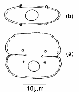
The semi-cells differ from the type in having rounded undulating margins with four very small granules within each margin and with a truncate or slightly convex apex, the walls are covered with pores. As with the type there are two prominent granules near the isthmus just above the deep closed sinus. and a single pyrenoid in the centre of each semi-cell. The figure below is (a) from the live sample, lower semi-cell in different focal plane to show pyrenoid, (b) from the preserved material, shows the end view of a semi-cell with the circular isthmus uppermost. Cells 20-23 um long; 24-26 um broad; 8-10 um thick; isthmus 7-7.5 um broad.
This is the first recording of this form from a British habitat, it is not in A Check-List of Desmids of the British Isles (1991).
Acknowledgements
The author is grateful to Mr. Alan Joyce for the samples from Scotland. To Dr. P.F.M. Coesel of the University of Amsterdam for his helpful comments, and to Dr. J.W.G. Lund for figures from The Fritsch Collection of Algal Illustrations (Freshwater Biological Association, England).
References
Brook A.J. and Williamson D.B. A Check-List of Desmids of the British Isles (1991) Freshwater Biological Association.
Coesel P.F.M. (1991) De Desmidium van Nederland. 4. (88pp). KNNV, Utrecht.
Gronblad R. (1921) Acta Soc. Fauna Flora. Fenn.49.
Huzel C. (1936) Beitrag zur Kenntnis der Mikrokopischen Pflanzenweit der Rauhen Wiese bei Böhmenkirchh. Veröffenlichungen der Wurttemberg Landesselle für Naturscutz. Heft 13 (pp 5-117).
Prescott G.W. Croasdale H.T. Vinyard W.C. & Bicudo C.E. De M. (1981) A Synopsis of North American Desmids Part II Section 3. University of Nebraska Press.
Note: 'um' in the text denotes micrometre.
Editor's note: Photomicrographs of the desmid and further notes on this article by the author Bill Ells were published in the May 1997 Edition of Micscape.
Comments and feedback via email to Bill Ells are welcomed.
Please report any Web problems
or offer general comments to the Micscape Editor,
via the contact on current Micscape Index.
Micscape is the on-line monthly
magazine of the Microscopy UK web
site at Microscopy-UK
Article archived at http://www.microscopy-uk.net/mag/art97/ellsnew.html
WIDTH=1