PHOTOMICROGRAPHY
by William Ells
Coniferae, Walnut Tree Lane, Loose, Maidstone, Kent, ME15 9RG,
UK.
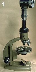 It is a fairly simple matter to take photographs
through a microscope, particularly if you have a microscope with
a fixed limb focussing, by raising and lowering the stage. My
first efforts were with a home made vertical tube on which to fix
the camera attachment plus the SL reflex camera with its lens
removed. A piece of brass tube from an old garden spray, a block
of wood to which was fixed a dovetail made of aluminium to fit
the microscope comprised the vertical tube. A much neater job can
be done if you have a lathe. (See the set up on a Vickers
microscope fig. 1).
It is a fairly simple matter to take photographs
through a microscope, particularly if you have a microscope with
a fixed limb focussing, by raising and lowering the stage. My
first efforts were with a home made vertical tube on which to fix
the camera attachment plus the SL reflex camera with its lens
removed. A piece of brass tube from an old garden spray, a block
of wood to which was fixed a dovetail made of aluminium to fit
the microscope comprised the vertical tube. A much neater job can
be done if you have a lathe. (See the set up on a Vickers
microscope fig. 1).
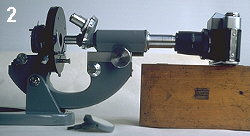 If you
have a microscope with fixed stage and focussing by raising and
lowering the tube the weight of the camera may push the tube
down, in this case you will have to support the camera on a
separate stand, or you could try using the microscope in a
horizontal position with the camera supported on something of
suitable height. (See fig.2).
If you
have a microscope with fixed stage and focussing by raising and
lowering the tube the weight of the camera may push the tube
down, in this case you will have to support the camera on a
separate stand, or you could try using the microscope in a
horizontal position with the camera supported on something of
suitable height. (See fig.2).
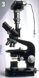
A much better arrangement than either of the
foregoing is a microscope fitted with a separate camera tube,
this has the advantage that the camera can remain in place while
viewing through the bino. or monocular, (see fig.3).
 If your objective lenses are parfocal you may be able
to switch from viewing to camera without adjustment, I have to
make minor adjustments viewing through the camera, here the
problems arise because the normal camera screen is unsuitable for
focussing highly magnified objects, you really need a plain
screen with cross wires. If using a cheap second-hand camera as I
do, a little luck is needed to get the best results.
If your objective lenses are parfocal you may be able
to switch from viewing to camera without adjustment, I have to
make minor adjustments viewing through the camera, here the
problems arise because the normal camera screen is unsuitable for
focussing highly magnified objects, you really need a plain
screen with cross wires. If using a cheap second-hand camera as I
do, a little luck is needed to get the best results.
If you are well endowed (financially), or have
had a grant for the purpose from a scientific society, you can
purchase a microscope with a built in camera system, ideally with
flash included and a good photograph is guaranteed every time.
Editor's note:
Three photomicrographs taken by Bill Ells of desmids, an
attractive green algae are shown below.
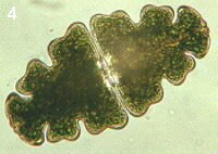 Fig.
4 Euastrum oblongum
Fig.
4 Euastrum oblongum
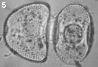 Fig. 5
Cosmarium botrytis. The semi-cells have come apart, it can
be seen that the end view is elliptical. The face view is that
normally seen. A reminder that desmids are three dimensional
objects, only a few desmids could be described as flat.
Fig. 5
Cosmarium botrytis. The semi-cells have come apart, it can
be seen that the end view is elliptical. The face view is that
normally seen. A reminder that desmids are three dimensional
objects, only a few desmids could be described as flat.
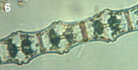 Fig. 6
Bambusina brebissonii
Fig. 6
Bambusina brebissonii
Editor's note
The Micscape Editor thanks William Ells for contributing this
article. Note that Bill Ells has written a number of articles on
desmids which are an attractive and fascinating algae, including
an Introduction
to the Desmids. All these articles can
be found in our Articles
Library in the Pond-life section.
© Microscopy UK or their
contributors.
Please report any Web problems
or offer general comments to the Micscape Editor,
via the contact on current Micscape Index.
Micscape is the on-line monthly
magazine of the Microscopy UK web
site at Microscopy-UK
WIDTH=1
© Onview.net Ltd, Microscopy-UK, and all contributors 1995 onwards. All rights
reserved. Main site is at www.microscopy-uk.org.uk with full mirror at www.microscopy-uk.net.