© R. Neumeyer 1997
© R. Neumeyer 1997
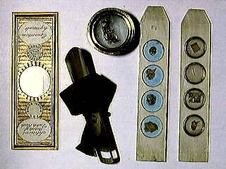
This picture illustrates several specimen holding
devices, the most recent having been made in the late Victorian
era. Beginning at the far right are two examples of ivory
"sliders" (hardwood was also used) dating from around
1800. The first has four holes drilled all the way through. Into
to each cavity a mica disc has been inserted (there is a shoulder
at one end). The specimen was placed on to the disc and a second
disc was secured on top by means of a small brass ring. This
slider was used for transmitted light observation.
Needless to say the mica slips are not perfectly clear so even with a modern microscope the final image is of poor quality, especially at high magnification. However, this slider and the one next to it, were in use at a time when microscope optics were greatly inferior to what is now available on a basic student microscope. For this reason mica slips did not present a serious problem. (In addition it was not possible for glass makers of the period to fabricate comparable slips). It was more common for microscopists of the time to view opaque specimens by means of reflected light, which is less effected by optical defects. The second slider is therefore more typical. Rather than holes, the manufacturer has drilled small cavities and the specimens were "glued" to the bottom.
The circular brass device at the top of the picture is an early "compressorium". It consists of a pair of short brass tubes, each with clear glass plate at one end. The subject, in this case an earwig, was placed inside the larger tube, then the slightly narrower tube was inserted and slowly pressed down onto the subject. The pressure between the top and bottom glass plates held the insect, or other live organisms stationary, allowing the microscopist to view it without unnecessary movement. (The squashed earwig in this particular compressorium has been there for almost 200 years)!
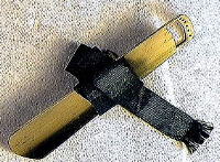 The long brass accessory in the centre of the picture
is a "fish plate". (Shown in more detail on the right
hand image). This one dates from around 1800, shortly after it
became widely known that blood corpuscles circulated within
vessels. Consequently, it was all the rage to place a small fish,
or frog, on this plate with the tail or foot webbing secured by
pins so that it stretched over the rectangular opening visible at
one end (the holes are for the pins). The device and it's
passenger were then mounted on the microscope stage with the
rectangular opening over the centre aperture. The microscopists
and guests then viewed blood corpuscles coursing through
capillaries in the clear skin of the webbing using transmitted
light from the substage mirror.
The long brass accessory in the centre of the picture
is a "fish plate". (Shown in more detail on the right
hand image). This one dates from around 1800, shortly after it
became widely known that blood corpuscles circulated within
vessels. Consequently, it was all the rage to place a small fish,
or frog, on this plate with the tail or foot webbing secured by
pins so that it stretched over the rectangular opening visible at
one end (the holes are for the pins). The device and it's
passenger were then mounted on the microscope stage with the
rectangular opening over the centre aperture. The microscopists
and guests then viewed blood corpuscles coursing through
capillaries in the clear skin of the webbing using transmitted
light from the substage mirror.
The last mounting device is more typical of present day preparations. However, it illustrates a peculiar habit of the Victorian microscopist (one of many!), namely the covering of mounted slides with paper! This particular example is relatively ornate, but by no means outstanding. Some of the paper covers are works of art in themselves, and are greatly sought after by collectors.
The Micscape and Microscopy UK Editors thank Ron Neumeyer for submitting this month's Image of the Month.
The author, Ron Neumeyer can be contacted by Email.
Please report any Web problems
or offer general comments to the Micscape Editor,
via the contact on current Micscape Index.
Micscape is the on-line monthly
magazine of the Microscopy UK web
site at Microscopy-UK