SAMWORTH'S SNIPPETS
Observation skills of the Victorians
by Mike Samworth
One thing that always amazes me when looking through my
favourite old microscopy books is the quality of the hand-drawn
plates. There is no better example of this than the plates in
'Micrographic Dictionary' by Griffiths and Henfrey. My copy is
the fourth edition, dating from 1883. Not only is this such
skilled draughtmanship but also a testament to their powers of
observation. It also illustrates the quality of the microscopes
they used all those years ago.
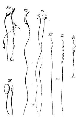
In the case of the plate shown, sperms from different
species, we only have to think of the size of these cells to
appreciate that we are talking about observations carried out at
high magnifications. It is interesting to speculate on how
accurate these diagrams are when compared with images taken on
more modern, sophisticated instruments today, and especially
since the advent of the electron microscope of course which has
contributed much to the study of sperm. The similarities and
differences between sperm of different species is something worth
contemplating. It is a sort of variation on a general theme and
the fact that the organisms can be so disimilar in other ways is
an example perhaps of both convergent and divergent evolution of
different parts within the same organism.
Snowflakes are dancing
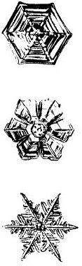
The Snowflake is almost recognised by everyone and epitomises
the beauty that is revealed when small objects are looked at more
closely. Indeed the Royal Microscopical Society has a snowflake
as it's emblem. Every snowflake has a unique history, and though
they all share the six-pointed symmetry, no two are alike.
It is the beauty and quality of the individual snowflake that
seems to attract our fascination. Knowledge of the hexagonal
character of snowflake crystals has a long history. Indeed there
in a book written by Han Ying in about 135 BC there is a clear
statement that 'flowers of snow are always six-pointed'. One of
the earliest European students was the 13th Century scholar
Albertus Magnus who commented on their star-like nature. Two
centuries later woodcuts were published that showed 23 varieties
of snowflake.
A well-known Scientist, Johannes Kepler wrote a lengthy study
in 1611 that not only perceived the hexagonal nature but also
tried to explain it on an atomistic basis. He was not wholly
successful, but his intuition was later proved true. Other famous
Scientists led their weight to further study, including Descartes
and the 17th century microscopist Robert Hooke.
Towards the end of the 19th century, the development of
photography opened up new possibilities for observation. A farmer
from Vermont, W A Bently, obtained some 5,000 pictures over 40
successive winters. Many of these were published in his 1931 book
'Snow Crystals'.
Ice crystals are formed from water droplets at certain
temperatures when they come into contact with 'nucleating agents'
such as tiny dust particles which trigger the crystallisation
process. So how can we explain their similarities and their
differences? Well, all water molecules form six-sided structures
as they freeze, and snow crystals are symmetrical because all six
sides are affected simultaneously by conditions in the cloud.
Growing outwards from the central dust particle, the unique shape
of each flake is determined by the shape of that particle and the
conditions under which the flake forms.
I was inspired to write this article by attending a lecture on
photomicrography of snowflakes recently and by the large amount
of snow we had just before and during Christmas this year. Why
not give it a go for yourself? For details of techniques contact
me Mike Samworth.
© Microscopy UK or their
contributors.
Please report any Web problems
or offer general comments to the Micscape Editor,
via the contact on current Micscape Index.
Micscape is the on-line monthly
magazine of the Microscopy UK web
site at Microscopy-UK
WIDTH=1
© Onview.net Ltd, Microscopy-UK, and all contributors 1995 onwards. All rights
reserved. Main site is at www.microscopy-uk.org.uk with full mirror at www.microscopy-uk.net.