Carchesium: an observation in pen and ink
by Wim van Egmond
Image © Wim van Egmond 1997
Carchesium: an observation in pen and ink
by Wim van Egmond
Image © Wim van Egmond 1997
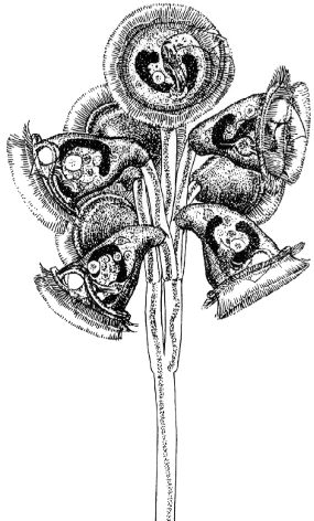
From a sample taken from a ditch near my home in Rotterdam I observed different kinds of vorticella-like creatures. Some had only one bell on a stalk (vorticella), others had more bells and formed a branching colony. I found several forms of these colonies. The way they contract can help determine the genus of the animals. Carchesium has spasmonemes (the inner structure of the stalk that enables it to contract) that are not connected so each bell's stalk can contract on its own. I made pencil sketches while looking at the animal from different sides with different lenses and lighting. The advantage of drawing instead of photographing is that you can combine several observations into one.
I made sketches of the interior organs (such as the sausage shaped macronucleus), the outline, the texture of the skin. Because the animal moves rapidly I had to make a quick sketch of the whole colony. The result of all this work was that I had a model-kit with which I could glue together the final drawing. I did this with pen and ink. What I found interesting was the way food-particles were transported by two rows of cilia into the opening on the side. An undulating membrane brought the food further into the body were it went into a droplet shaped food vacuole.
One of the things I noticed after comparing my drawings to other peoples drawings was that nobody pictured the undulating membrane protruding out of the mouth-opening. When observing it carefully I noticed it sticking out in the form of a translucent sheet. Can anyone confirm this?
The interesting thing about drawing microscopic organisms, and you don't need any special device for that except a pencil, paper and a keen eye, is that you learn far more than by making photographic pictures. After making the drawings you are also more able to recognize different kinds of organisms.
Note about the artist.
Wim van Egmond (born 1966) is a professional artist and
photographer who is very interested in microscopy and natural history.
His specialization is 3D photography, also through the microscope.
Editor's notes.
The author Wim
van Egmond I'm sure would welcome comments.
Note that the image should not be used without first
seeking the permission of the artist.
The Microscopy UK and Micscape Editors would like to thank Wim for sending this tremendous drawing. Also thanks to Jan Parmentier for liaising with Wim to send us this material and for Anne Bruce for scanning the image. The drawing was first published in 'Microwereld' the magazine of the Dutch Society for Microscopy.
Micscape is the on-line monthly magazine of the Microscopy
UK web
site at Microscopy-UK