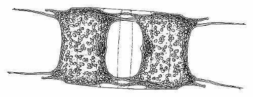
Biddulphia sinensis
Marine Diatoms
by Jan Parmentier
A diatom is in essence a pill box of glass; the outer wall of the cell is of hydrated silica with an inner pectic layer. We can find diatoms where there is water, but it is especially rewarding to study them in the sea. You can study marine diatoms during the entire year, but really spectacular results will be obtained in the spring (March / April) and in the autumn (September).
In temperate waters nutrients accumulate during the winter. This results in a spring bloom of phytoplankton. Sufficient light is available then and temperatures are higher. The most important factor however is the availability of nutrients. The spring outburst of phytoplankton is grazed down by zooplankton in the summer and occurs again as a second maximum in the autumn.
Due to the death and decay of the spring plankton, fresh nutrients become available in the autumn. In tropical seas there is always a scarcity of nutrients; there we don't see such spectacular blooms and in cold, northern seas there is only one bloom a year. Of course, this is a generality and the occurrence of primary and secondary plankton blooms depends on the exact geographical and climatic factors.
For diatoms, silica is especially important and a limiting factor. In the Netherlands, the Oosterschelde is a favourite place for our club to collect diatoms. We can find in our Northern seas about 160 different sorts of planktonic diatoms, of which 112 are of the round type and the rest of the more elongate type. So we have two classes of diatoms: the Centricae (round forms) and the Pennatae (elongate) forms.
Reproduction occurs mostly asexually. A round diatom is a sort of pill box. Prior to division, two new box ends are formed in the middle of the cell. Separation then occurs and as a result, one of the new cells has the same size as the original cell and the other new cell is smaller than the original cell. This could result in cells getting smaller and smaller, but at a certain size the diatoms regain their size by what we call auxospore formation.
Biddulphia sinensis
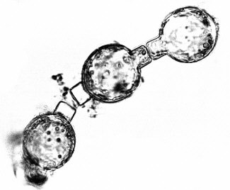 Auxospore formation in Centricae is a method of sexual
reproduction. After fusion of egg and sperm cells a zygote is
formed, which enlarges to an auxospore, a large, spherical cell.
In photo 1 (on the right) we see auxospores of Melosira.
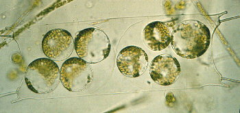
From the auxospores, normal diatoms are formed. The male cells
are formed in spermatogonia, to be seen in photo2 (on the left
below) in the species Biddulphia sinensis.
Auxospore formation in Centricae is a method of sexual
reproduction. After fusion of egg and sperm cells a zygote is
formed, which enlarges to an auxospore, a large, spherical cell.
In photo 1 (on the right) we see auxospores of Melosira.
From the auxospores, normal diatoms are formed. The male cells
are formed in spermatogonia, to be seen in photo2 (on the left
below) in the species Biddulphia sinensis.
Sexual reproduction in the Pennatae, the more elongate type of diatoms, occurs by male and female gametes that look equal. The first microscopists were so fascinated by the beautiful glass cell walls with wonderful marks and perforations that the study of the biology and reproduction came much later than the phase of description. To be honest, to study the reproduction, it is necessary to grow diatoms in the laboratory, a difficult task. In material collected in nature however, we sometimes see spermatogonia and auxospores.
 Floating diatoms, in order to survive, must stay in the
upper, illuminated layer of the sea. So they possess several
adaptations to keep them near the surface. Planktonic diatoms
have in general lightly built silica cell walls, different from
the more heavier built sessile marine species.
Floating diatoms, in order to survive, must stay in the
upper, illuminated layer of the sea. So they possess several
adaptations to keep them near the surface. Planktonic diatoms
have in general lightly built silica cell walls, different from
the more heavier built sessile marine species.
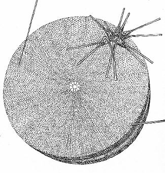 Coscinnodiscus concinnus is a large round pill
box, with a large vacuole, filled with cell sap of the same
density as sea water (shown on the right with Asterionella
glacialis). Other diatoms are very long and thin, like a
pencil. Some species are united in long chains, by hairs or
threads. In other species, each cell has four long horns, to
increase the surface resistance offered to the water. Apart from
helping in staying afloat, these devices may also serve as a
defense against grazing by zooplankton.
Coscinnodiscus concinnus is a large round pill
box, with a large vacuole, filled with cell sap of the same
density as sea water (shown on the right with Asterionella
glacialis). Other diatoms are very long and thin, like a
pencil. Some species are united in long chains, by hairs or
threads. In other species, each cell has four long horns, to
increase the surface resistance offered to the water. Apart from
helping in staying afloat, these devices may also serve as a
defense against grazing by zooplankton.
In the pictures you will see examples of these several adaptations. When you are collecting diatoms with a plankton net you will also find quite an assortment of animal species belonging to the zooplankton. Copepods, larvae of echinoderms, crustaceans, polychaetes, molluscs, coelenterates and many others can be found and are all most interesting for microscopists!
Editor's notes:
Comments to the author Jan Parmentier (Chairman of
the Dutch
Society for Microscopy) are welcomed.
The photographs are by Jan Parmentier and the drawings by Wim van
Egmond.
Jan Parmentier and Wim van Egmond have provided Micscape with a wealth of beautiful photographs and drawings of marine diatoms. Micscape will be presenting these in a Marine Diatom gallery in the near future.
Please report any Web problems
or offer general comments to the Micscape Editor,
via the contact on current Micscape Index.
Micscape is the on-line monthly
magazine of the Microscopy UK web
site at Microscopy-UK