There was a time long ago when advertisements for toothpaste told an unfortunate truth: brush with Brand X, and your teeth were guaranteed to shine as never before. An explanation was never given- for good reason. Vigorous brushing with Brand X scoured your tooth enamel as thoroughly as sandpaper! When I got my first microscope in the 1930s, I learned that if you took Brand X (there were others with the same ingredients), squeezed some toothpaste and diluted it with a little water, then placed a drop on a slide and examined it under a microscope, you would enter a glassy wonderland of beautiful geometric shapes. They were the ornamented siliceous shells of diatoms.
Diatoms first appeared 200,000 million years ago, but flourished in Mesozoic times (ten million years ago) when they lived in such enormous numbers they left vast deposits of their shells up to three thousand feet thick. We call this diatomaceous earth and use it for filters, insulation, and as an abrasive. That last quality appealed to toothpaste manufacturers. What did they care if you wore your teeth down to nubs, as long as they shone? Eventually the government stepped in, the practice was deemed injurious and for the microscopist an easy source of wonderful shapes was gone.
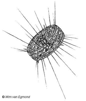
Over the years I've seen living diatoms in oceans, rivers, lakes and in the soil. They never fail to astonish. Although a diatom is a microscopic plant, it can grow in huge quantities until the resulting mass becomes visible to the naked eye. It is the intricate architecture of an individual diatom that is so fascinating.
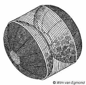
The glassy shell of a diatom comes in two parts, one fitting over the other exactly like a container with a lid. The upper and lower halves are sculpted with exceedingly fine lines that actually are rows of ultra-microscopic pores through which the enclosed protoplasm remains in touch with the exterior environment. There was a time after the Civil War when microscope manufacturers were vying with one another to produce the highest quality lenses, but in an age without computers or a means of etching micrometer rulers, they turned to the one microscopic object that was perfection. Diatoms were placed under a new instrument and if their decorative lines appeared perfectly parallel or symmetrical, the technician had succeeded in making a distortion-free lens. But if the lines were wavy or fuzzy, the lens-which may have taken months to grind and mount in a metal objective-was summarily chucked out. Nature was perfect, human efforts were not.
The rigid pillbox shell of a diatom suggests a question: how does the cell reproduce? After the internal cellular contents bunch together in preparation for cell division, the lid comes off and half of the protoplasm forms a new bottom, while the bottom-now serving as a lid-forms a new bottom. Each of the two offspring therefore have one of their parent's shells. Think about that. With every generation one of the two daughter diatoms is going to be progressively smaller than its parent, and if this went on forever, they would simply vanish into nothingness. Obviously such reduction doesn't continue for long. From time to time a daughter cell discards its single parental shell and produces two new larger ones, returning to its ancestral size in one fell swoop.
The asexual reproductive process (far more common among diatoms than sexual reproduction) occurs mostly after dark and can result in enormous numbers over a short period-a billion in a month from a single ancestral diatom. Why aren't oceans and lakes clogged with diatoms? Despite their hard glassy shells, they are a favorite food for countless kinds of small animals. Watch a pond snail rasp its way across a submerged rock; using its file-like tongue to remove blankets of diatoms. The glass sides of an aquarium often develop a splotchy brownish coating of diatoms. Put in a couple of snails and they will happily browse through the unsightly stain, leaving clear zigzag paths behind where the coating of diatoms has been scraped bare by the snails' tongues.
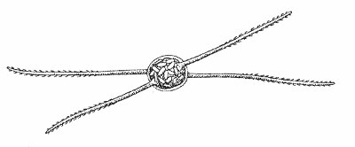
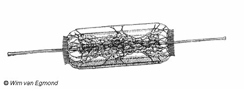
The variety of diatom shapes seems endless, although individuals of each species are identical to one another, except for size. What surprises a watcher of diatoms is the movement of some, slow or rapid depending upon the species. If you watch a long, spindle-shaped diatom, it moves across the microscope's field of view in slow and stately fashion. Suddenly it reverses itself and heads back where it came from. Movement is made possible by slender bands of cytoplasm streaming out from clefts in the lid and bottom. The strands flow out, then in circuitous fashion go back into the shells again. They remind me of endless caterpillar tractor treads. Movement obviously benefits these tiny plants, allowing them to congregate where sunlight enters the water.
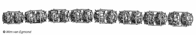
One needlelike species congregates in a flat stack of individuals parallel to one another. As you watch, the entire array suddenly zips out, expanding into a very long chain that resembles an old-fashioned carpenter's rule. After being extended for a moment, the colony slides back, either back into its original parallel array, or it keeps on until all individuals are extended out as far as they can go in the opposite direction. Somehow each cell "communicates" with its two adjacent neighbors, but so quickly it appears the entire colony acts simultaneously.
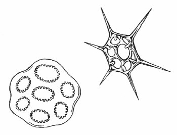
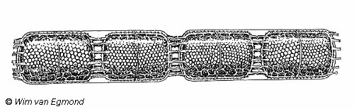
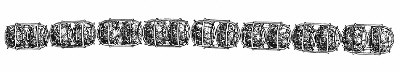
There are over five thousand species of diatoms. Some move, some don't. Some are circular, others are elongated, or square or rectangular or shaped like dumbbells. Some colonies resemble jointed dominoes. Because of the great variation of shape and the sculpturing of their shells, they have fascinated microscopists and botanists for centuries. There still are scientists who spend their lives studying these fascinating little plants with good reason, because diatoms are superb indicators of water quality.
Other biologists seek new species of diatoms. I visited Paul Conger at the Smithsonian Institution many years ago (he was a distant relative of my wife), but to do so we had to climb up one of those ancient red brick towers to his laboratory at the very top where we could look over the entire Mall. The room was crowded with shelves reaching the ceiling, each shelf holding hundreds of microscope slide boxes, every one of which contained a hundred slides he had mounted or acquired.
Long ago when they occasionally got bored doing whatever they were doing with diatoms, microscopists would select certain ones under a microscope, then move them about on the slide with a single human hair mounted on a wooden shaft. Imagination ran riot and they created geometric designs, pretty scenes and cartoons before cementing them in place. From his collection Paul pulled out some of his prizes and showed them to us, several over one hundred years old. There under a microscope were ornamental designs and mosaic landscapes of mountains and trees and birds.
A few showed people dancing or throwing balls to running dogs. Once I clumsily tried moving some diatoms around on a slide, but they flipped off into space and vanished. I couldn't even get three or four lined up near one another. Yet two centuries ago men and women with greater patience and skill created intricate arrangements from diatom shells, then preserved the tiny scenes for others to see. It was only a sidelight and not science, but it must have been fun. I wonder if there is anyone in the world today bothering with such artistry that only a privileged few might ever see.
© 1995 William H. Amos
Editor's notes:
Comments to the author Bill Amos are welcomed or to the illustrator Wim van Egmond.