SOME NOTES ON THE DESMID
GENUS GENICULARIA
William Ells
Coniferae, Walnut Tree Lane, Loose, Maidstone, Kent. ME15 9RG.
- Phylum Chlorophyta
- Class Zygnemaphyceae (formerly
Conjugatophyceae)
- Order Desmidiales (Placodermae)
- Sub order Closteriinae (Archidesmidiinae.
Lind & Brook 1980)
- Family Gonatozygaceae
- Genus Genicularia De Bary 1858
- Species Two species found in Britain G.spirotaenia
& G.elegans.
Genicularia are tubular cells without
a median constriction, the cell wall is covered with minute
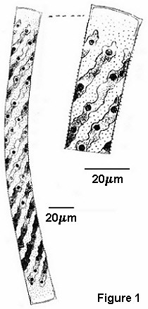
granules, ( W.&.G.S. West (1904) (1) figure G.spirotaenia
from Loch Beosetter in the Shetland Islands, Scotland, with
minute spines Fig.1). They were originally classed with
the Saccoderm desmids but now together with Gonatozygon
they have been transferred to the Placodermae, minute pores
having been found in the walls of the related genus Gonatozygon,
Genicularia are assumed to have the same wall structure.
 " The genus Genicularia is one of the
rarest of all known genera of Desmids" W.& G.S.West
(1904) (2). The author has found the genus in samples from two
sites on the Isle of Skye, Glen Drynock, and Loch More-na
Caiplaick, Scotland collected by Mr Alan Joyce, who states the
genus is variable and not uncommon in the Rhiconich area of
Sutherland, Scotland.
" The genus Genicularia is one of the
rarest of all known genera of Desmids" W.& G.S.West
(1904) (2). The author has found the genus in samples from two
sites on the Isle of Skye, Glen Drynock, and Loch More-na
Caiplaick, Scotland collected by Mr Alan Joyce, who states the
genus is variable and not uncommon in the Rhiconich area of
Sutherland, Scotland.
The size range given by W.&.G.S.West (2) is
:- G.spirotaenia De Bary 1858 cells 20-25 µm broad,
200-400 µm long. Length - Breadth 10-20 µm. G.elegans
W.& G.S.West (1903), is a more slender sp. 14-16.3 µm broad,
303- 427 µm long. Length -Breadth 21-30 µm. The size range in
the sample from Skye is :- 16-18.5 µm broad, 222.5-300 µm long,
Length - Breadth 14-18.5 µm slightly narrower than the sizes
given for G.spirotaenia above, most of the cells were of
greater breadth than that given for G.elegans, 12 cells
were measured 2 filaments of 3 cells and 6 individual cells.
Figure 1. Complete cell and enlargement to show spines
There are two parietal spiral chloroplast in each cell from the
Isle of Skye with 2 to 6 'turns' in each cell. Some showing lax
turns and others tighter with more turns, photo' shows part of
cell. W.& G.S.West(2) figure G.elegans with laxer
spirals than G.spirotaenia Fig.2. Contradicted by
Prescott et al (1972) in their text, G.spirotaenia:-
'more lax turns than G.elegans.'
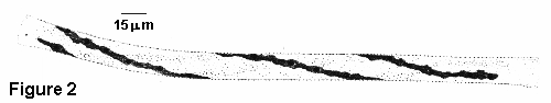
Figure 2. Complete cell
Observations :-
Filaments of 3 cells joined, and individual cells, some
geniculate (they bend like a knee joint to conjugate),
distinguished from the related filamentous algae Spirogyra
by the slightly enlarged apices of each cell, the small granules
which make the cell walls appear rough and the fact that the
cells readily disassociate when attempts are made to manipulate
them. Although there is no median constriction of the cell wall,
or median division of the chloroplast; that is, they are not
obviously semi-cells; as in most of the placoderm desmids, it
could be seen in some individual cells that there are two
semi-cells as one is distinctly more mature than the other, the
granules being slightly larger and clearly seen, the younger
semi-cell appearing smooth by comparison. The cells with 6 turns
of the 2 intertwined chloroplast had 3 turns in each semi-cell
joined by a short straight section about the median area, thus
the chloroplast although continuous was clearly divided between
the two semi-cells. Indian ink added to the water showed up a
mucilage envelope, this mucilage appeared thin and absorbed some
of the ink, showing as a lighter grey rather than white like the
desmids with a copious mucilage.
During later observations one was cell found
344.5 µm x 11 µm only vestiges of chloroplast could be seen,
size suggest G.elegans. Other specimens were seen with a
slight swelling of the cell walls as though new apices were
forming in the median area and the chloroplast was also dividing
at this point. Many desmids will live and reproduce for months,
even years, if kept in a cool place in North light, but the Genicularia
chloroplast deteriorated within a few days.
Williamson (1992) in a comprehensive survey of
the desmids of the Shetland Isles, collected from Loch Beosetter,
(where the West's recorded G.spirotaenia) no Genicularia
were recorded.
Conclusions:-
Although there is some variability in the laxity or otherwise of
the chloroplast spirals and the breadth of most cells were larger
than those recorded by W.&.G.S.West for the species, the
specimens from Skye were considered to be G.elegans.
There are filaments of Gonatozygon
aculeatum Ruzicka in the samples, typically 203 x 8 µm,
single spiral chloroplast, short spines on cell walls.
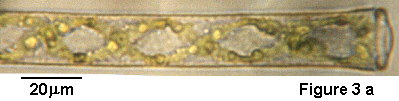
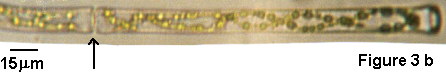
Figure 3a and 3b. Photomicrographs, arrow points to where
cells join.
Acknowledgements:-
The author is grateful to Mr. Alan Joyce for
the samples and his helpful comments. To Dr. J.W. Lund curator of
The Fritsch Collection of Alga Illustrations at The Freshwater
Biological Association, and to Mr. David Williamson for a copy of
his paper on the desmids from the Shetland Isles. References:-
Lind E.M. Brook A.J. 1980. Desmids of the
English Lake District, Freshwater Biological Association,
Scientific Publication No.42.
Prescott G.W., Croasdale Hannah T.,&
Vinyard W.C. 1972. North American Flora. Series II. Part
6.
West W.&.G.S. 1904 (1). Freshwater
Algae from the Orkneys & Shetlands. From The
Transactions & Proceedings of the Botanical Society of
Edinburgh. Ed. D.J.Scourfield.
West W.&.G.S. 1904 (2). A Monograph of
the British Desmidiaceae. Vol.1. Ray Society.
Williamson D.B. 1992, A Contribution to our
Knowledge of the Desmid Flora of the Shetland Islands, The
Botanical Journal of Scotland.
Editor's note: Comments and
feedback via email to Bill Ells are welcomed, and will be passed on to the author
William Ells. Or contact the author directly via the postal
address above.
© Microscopy UK or their
contributors.
First published in Micscape Magazine, August
1997 ( ISSN 1365 - 070x )
Article archived at
http://www.microscopy-uk.net/mag/art97b/ellsgen.html
Please report any Web problems
or offer general comments to the Micscape Editor,
via the contact on current Micscape Index.
Micscape is the on-line monthly
magazine of the Microscopy UK web
site at Microscopy-UK
WIDTH=1
© Onview.net Ltd, Microscopy-UK, and all contributors 1995 onwards. All rights
reserved. Main site is at www.microscopy-uk.org.uk with full mirror at www.microscopy-uk.net.