Playing Detectives
(A
bit of fun but a learning experience too!) by Maurice
Smith. Aug. 1997
What isn't about Microscopy?
I recently had my leg pulled over one of our
front page animation's. The one concerned was of two crickets (or
are they locusts?) running along piggy-backed as they embarked on
their favourite past-time of making more crickets. "It made
me laugh...", said my friendly tormentor: "...but what
has it to do with microscopy?"
This is the not the first time such a question
has been asked of the crew here at Micscape and Microscopy-UK.
The problem here lies with different perceptions about what
Amateur Microscopy means. Is it for example about studying life
and processes only at the microscopical scale, or is it about
studying life in general along with its processes - and using
microscopy as one tool of many to help reveal greater truth and
insight?!
I believe everything there is - man, beast,
cosmos, nature... you name it, I'll include it too - is
built upon the foundation of microscopical processes. So this
month, I thought I'd demonstrate to you just one such connection
between the everyday world and a process in the microscopical
one. We are going to play a little game together: a game
where we become Amateur Microscopy Detectives!
A Question...
What is the connection between
the diagram below and a criminal act?
Okay, I guess it's not a very good diagram but we
will take a look at a better one later. Maybe, you'll get to see
the connection between biology, microscopy, and criminal acts if
I hold up a bit of my anatomy for you to see.... NO... not that! This is a family
site! Here's the bit I meant... 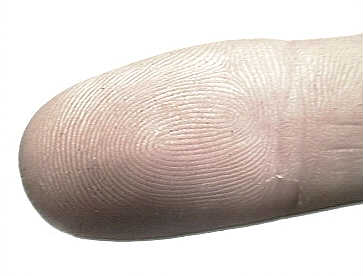
YUP! It's my finger tip, or more precisely -
the fine epidermal ridges which comprise what is commonly called
: a finger-print! You start to see the connection now with crime,
I bet. We all have a unique pattern of lines like this on our
finger tips. Normally, they fall into three categories: arch,
loop, and whorls, although variations between loops and whorls
can exist. I'm not an expert but I think my one is a
double-whorl. Their names are self explanatory but you may need
to look very carefully to distinguish between loops and whorls.
In the UK, loops are commonest (70% of the
population), whorls are next (25%), and arches are the rarest at
5% of the population. Take a look at your one now - what type is
it?
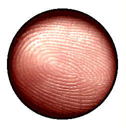
Not easy to see, eh..? Well
get out your trusty 8x magnifier and take a look:-
 Most people know
that many crimes have been solved though the use of a technique
called 'finger-printing'. When someone commits a crime,
especially where it wasn't planned, they will inevitably have
touched something at the crime scene. In doing so, they will have
left behind an invisible mark which can be proven to have
belonged to only one human-being on the planet. Special people
visit a crime scene with the police. By dusting objects with a
fine powder, they are able to reveal finger-prints
left behind....
Most people know
that many crimes have been solved though the use of a technique
called 'finger-printing'. When someone commits a crime,
especially where it wasn't planned, they will inevitably have
touched something at the crime scene. In doing so, they will have
left behind an invisible mark which can be proven to have
belonged to only one human-being on the planet. Special people
visit a crime scene with the police. By dusting objects with a
fine powder, they are able to reveal finger-prints
left behind....
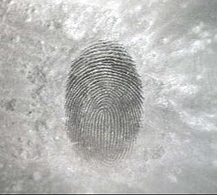 ...
like my one here!
...
like my one here!
There's no magic involved. The chalk dust that
I used to get this print works just the same way as the finer
powder used by the people investigating crime-scenes: the dust
sticks to the fine lines of sweat which have been deposited on
the object from the high points (ridges) of the suspect's skin.
Where the lines are dark, no sweat was deposited because it was
still trapped in the low points (valleys) of the skin.
Can you imagine all the crimes
that have been solved simply because we possess two
biological features: patterns on the surface of our skin,
and sweat glands!
Now, as it happens, sweat glands
in the human skin are quite difficult to spot in specimen slides.
Many students and amateur microscopists have to play at being
detectives themselves to find them. Here, let me show you a
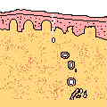
rather bad specimen of a skin section. This one below was
borrowed from a school where biology students would have been
using it to identify processes like sweat glands along with a lot
of other things present in human skin.

I've marked two areas of interest as A
and B which we I will be referring to in a moment, but first
of all, let's take a look at a diagram from a typical biology
text book and see how it helps (huh... I wonder about some of
these books!) us to identify where the glands are what we should
be looking for.
Here's a very quick and dirty diagram. Text
books normally show skin sections more refined than this.
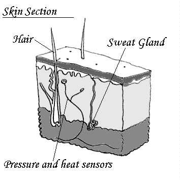 That wormy,
wrigley, looking thing is the process we wish to consider in more
detail. The problem is that this 3D solid looking section of skin
has been put together conceptually through the mind of an artist.
In real life, to see a skin section, you have to slice it up so
thin that you only get to see bits of the items (processes)
buried in the skin. If you are lucky, the section you get in the
slide to study might contain a bit of everything. It is rare to
get perfect examples of all - or even the majority of things you
would wish to study - in a single section!
That wormy,
wrigley, looking thing is the process we wish to consider in more
detail. The problem is that this 3D solid looking section of skin
has been put together conceptually through the mind of an artist.
In real life, to see a skin section, you have to slice it up so
thin that you only get to see bits of the items (processes)
buried in the skin. If you are lucky, the section you get in the
slide to study might contain a bit of everything. It is rare to
get perfect examples of all - or even the majority of things you
would wish to study - in a single section!
So how the heck do you identify a sweat gland.
Well you have to follow a few clues. First of all - see how the
gland starts out to be very entwined at its base and then twists
and spirals up to the surface of the skin, like a winding tube
getting thinner and thinner?
You do... good! Let me show you a slide where
that 'wrigley' bit at the base is present:-
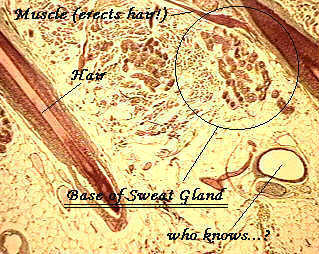 What you
actually get to see is a pattern of circles and elongated
circles: cross-sections of the sweat gland tube in many
places as it loops back and forth, around and around!
With a little care, you can even get to see which circles
represent sections of duct as distinct from secretory parts of
the gland.
What you
actually get to see is a pattern of circles and elongated
circles: cross-sections of the sweat gland tube in many
places as it loops back and forth, around and around!
With a little care, you can even get to see which circles
represent sections of duct as distinct from secretory parts of
the gland.
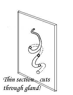
The duct itself 'meanders' up to the skin
surface. The tube gets thinner, and a section through it yields
small circles (or rings) representing the duct wall. Here's
another diagram (very quickly done) to demonstrate:-
 After looking at
this, go back up and examine the image with A and B indicating two
processes. I reckon that A is not a duct because even though it 'wanders' up to the
surface, it is intact: more likely to be muscle involved with
erecting body hair! Item B does look like a trail of tiny circles, maybe a sweat
duct, very thin, as it approaches the skin's surface. But
there is not the 'tell-tale' system of clustered circles at the
end of the trail... so is it a sweat gland duct or one of many
other processes just beneath the surface of the skin? I don't
know either!
After looking at
this, go back up and examine the image with A and B indicating two
processes. I reckon that A is not a duct because even though it 'wanders' up to the
surface, it is intact: more likely to be muscle involved with
erecting body hair! Item B does look like a trail of tiny circles, maybe a sweat
duct, very thin, as it approaches the skin's surface. But
there is not the 'tell-tale' system of clustered circles at the
end of the trail... so is it a sweat gland duct or one of many
other processes just beneath the surface of the skin? I don't
know either!
It's a heat wave here in the UK. A hot
and humid night. I have a cooler on in in my room to keep down
the heat from computers, lights, cameras, and the extraordinary
weather. My body is fighting to evaporate moisture to maintain a
constant temperature, as I struggle to make ready this article.
If my sweat glands packed up... where would I be?! The humidity
is high, the sweat remains like a thin film on my body. I am
growing confused... where did I begin this page.... ah... yes:
crime, diagrams, and connections!
Go back up to the very first image at the top
of the page. Can you see the connection now? If so, you have
taken your first step into becoming a Microscopist detective. Welcome to a whole new world!
Maurice Smith - August 1997 -
"Sweating like mad!"
Comments on the article to the author Mol (Maurice Smith)
© Microscopy UK or their
contributors.
Please report any Web problems
or offer general comments to the Micscape Editor,
via the contact on current Micscape Index.
Micscape is the on-line monthly
magazine of the Microscopy UK web
site at Microscopy-UK
WIDTH=1
© Onview.net Ltd, Microscopy-UK, and all contributors 1995 onwards. All rights
reserved. Main site is at www.microscopy-uk.org.uk with full mirror at www.microscopy-uk.net.