Contrast
by Mike Samworth
Contrast is very important in microscopy if detail is to be
seen in the subject. Some would argue that it is as equally
important as resolution. Contrast in the specimen can be achieved
in a number of ways. Some are treatments of the subject itself,
such as staining, others are optical methods used during the use
of the instrument. To illustrate these differing methods two
photomicrographs are shown.
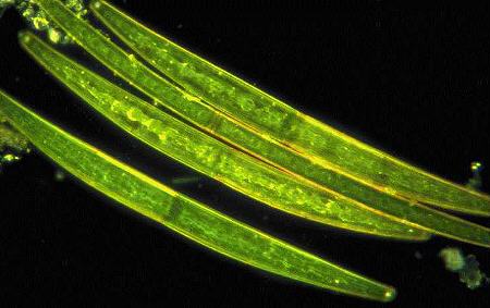
This first photomicrograph shows the desmid Closterium,
one that many will be familiar with. To increase contrast
dark-field illumination has been employed. Though in this case it
is probably done for purely artistic reasons, this type of
illumination can make more visible structures that are very
difficult to make out otherwise. A useful analogy to use is that
of not being able to see small dust particles in a well-lit room,
but them being easily visible when lit by a shaft of sunlight
coming through a window into a darkened room.
Closterium.
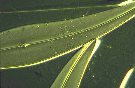
The second method of contrast I have chosen to
illustrate is of the specimen treatment type. In this case the
specimen is that well-known diatom Pleurosigma angulatum.
This diatom is a much-used test object for checking the
resolution of objectives. Using a x40 objective the punctae
should be resolved as dots, not lines, if the microscope is set
up properly. Unfortunately, the frustule is so pale that it is
hardly visible, even when mounted in a substance of high
refractive index. In the photomicrograph shown, the diatoms
visibility has been enhanced by being coated in aluminium, a
technique perfected by the late Horace Dall. It is worth noting
that although the dots are visible in the original transparency,
at the sort of resolution achieved through scanning and
subsequent display on your computer screen, they are no longer
so!
Pleurosigma angulatum,
aluminium coated.
Both photomicrographs by Mike Samworth.
If any reader wishes to ask about any of the
above, or to comment, please do get in touch by contacting me Mike Samworth
© Microscopy UK or their
contributors.
Please report any Web problems
or offer general comments to the Micscape Editor,
via the contact on current Micscape Index.
Micscape is the on-line monthly
magazine of the Microscopy UK web
site at Microscopy-UK
WIDTH=1
© Onview.net Ltd, Microscopy-UK, and all contributors 1995 onwards. All rights
reserved. Main site is at www.microscopy-uk.org.uk with full mirror at www.microscopy-uk.net.