The late G. T. Harris (1928) Hon. Member QMC.
wrote 'one variety of E. crassum (fig. 1) is quite
distinctive, and it is really remarkable that such a well marked
form should have escaped being named as a variety when so many
ill-defined and slightly variable forms have been given varietal
rank. It will be seen that two prominent projections occur in the
middle of the semi-cell outline. Usually only one semi-cell in
each plant has the variation, but I have plants with both
semi-cells having the variation, as in the figure drawn. In such
cases it makes a very distinctive looking plant'.
I could not agree more and his remarks apply to other forms difficult to tell apart that are given specific rank, although the morphology of the cell is not the only criteria determining species and sub-species; the chloroplast and/or number and disposition of the pyrenoids and/or morphology of the zygospore may differ. Though I am not aware of any zygospores having been recorded for E. crassum.
The genus Euastrum differs from other genera of desmids with one exception in having a usually deep cleft or notch in the apices, although in some species E. pectinatum for example it is a mere depression, and in others only a very small notch. The exception is the genus Tetmemorus which also has a deep cleft in each apex, however the cells are cylindrical whereas in Euastrum the cells are somewhat flattened or elliptical in end view. Harris gives the measurements for E. crassum as:- breadth 80 - 106 micrometres, length 160 - 200 micrometres which agrees with my own observations.
The specimen figured is a good example of those forms that are markedly different but unnamed. The habitat is not given. Harris lived in Devon and collected mainly from his home county and Dorset. I have a figure from the Fritsch Collection of Algae Illustrations of Euastrum crassum (Breb) Kutz. by Allorge 1930 showing the two projections on both semi-cells. Williamson (personal communication) informs me he has collected this form from Boo Tarn in the Lake District and from Thursley Common Surrey. I have found it in two samples sent to me by Mr Alan Joyce, one from a site in Sutherland and another from the Isle of Skye, (fig. 1) the latter containing several cells. It is therefore widespread in its occurrence and has appeared over a period of more than sixty years, it cannot be dismissed as a mere anomaly.
Although Harris describes this form as unnamed, in the index to the figures he gives the form the epithet mamillatum thus:- Euastrum crassum var. mamillatum mihi. There is no reference to this in his text.
Mr Harris's suggestion does not appear to have been taken up and the form remains unnamed in the literature, possibly because he does not give a description in Latin, perhaps forma often used as a lower rank of variety would be more appropriate. There does not appear to be any universally accepted rules as to when variety, forma or facies is to be used in the description of an intra-specific form.
Mr Harris's figure of E. crassum (Breb) Kutz. (fig. 2) narrows towards the apices in a manner that I have never seen in any collection and I have not seen figured elsewhere. My fig. 3 shows the form of cell usually seen.
In conclusion it is interesting to consider the specimens with the nodules on one semi-cell only. I have not found any, they are recorded by the West's (1905) by Harris (above) and by Dick (1930). How did they arise? Perhaps by the conjugation of a type (fig. 3) specimen with a specimen of the mamillate form. Aberrant and asymmetrical forms can be produced in other genera when conditions change during division, that is the daughter cells do not develop fully Brook (1981). And what happens when a specimen with the nodules/mamilla on one semi-cell only divides? Until specimens are found and division observed one can only speculate.
Acknowledgements: Thanks to Dr. J. Lund for figures from the Fritsch Collection of Algae Illustrations, to Mr. A. Joyce for samples from N. W. Scotland. To Mr. D. B. Williamson for information on habitats, and to Mr. Colin Lamb for a copy of Mr. Harris's article from Watsons Microscope Record.
References.
Brook A. J. (1981). The Biology of Desmids. Blackwell Scientific
Publications.
Harris G. T. (1928). The Desmidiacea. Chapter VI, pages 8-9,
plate V, figs. 1 & 2. Watsons Microscope Record. No. 14 May
1928.
West W. & G. S. (1905). A Monograph of the British
Desmidiaceae, Vol. 2. Ray Society.
Dick (1930). Figure from the Fritsch Collection of Algae
Illustrations.
Author's Note: Since this paper was published Mr. Barry Ellam has sent me a photo-copy of a paper by Professor Lenzenweger that appeared in 'Mikrokosmos' No 83. Mai 1994. On page 156 is a photomicrograph of Euastrum crassum with two nodules on each semi-cell. Thus the form is to be found in Central Europe as well as Great Britain.
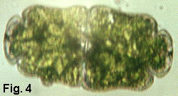
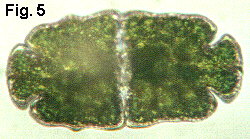
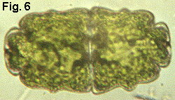
Recently (October 1997) I examined a sample of water from a bog on Dartmoor, in the County of Devon, England. In this sample were several specimens of E.crassum. Fig.4. is a photomicrograph of a cell with a nodule on one semi-cell. Fig 5. with two nodules on one semi-cell. Fig 6. is a cell with two nodules on both semi-cells from the original sample from Sutherland, Scotland.
Editor's note
The Micscape Editor thanks William Ells for contributing this
article, which first appeared in the Bulletin of the Quekett
Microscopical Club. Note that Bill Ells
has written a number of articles on desmids which are an
attractive and fascinating algae, including an Introduction to
the Desmids. All these articles can be
found in our Articles Library in the
Pond-life section.
Comments and feedback to the author William Ells welcomed.
Please report any Web problems
or offer general comments to the Micscape Editor,
via the contact on current Micscape Index.
Micscape is the on-line monthly
magazine of the Microscopy UK web
site at Microscopy-UK
Article at http://www.microscopy-uk.net/mag/art98/ellsecra.html
WIDTH=1
![]()