Photographing
micro-organisms
by
Steve Durr
(Page Design by Anne Bruce April 1998)
Epping Forest
abounds with small ponds and dikes and is therefore an excellent
place to find freshwater protozoa and algae. I use a round net
with two filters of 53 micrometers and 35 micrometers, these two
filters hold all the specimens that I need. It is important to
make sure that the pond water is placed into a suitable container
and kept fairly cool, especially on very warm summer days, where
heat will very quickly kill off many species of protozoa.
Many of the
micro-organisms found in ponds are very difficult to observe in
any detail let alone trying to photograph them. One way to
increase the contrast when using brightfield illumination is to
close down the substage condenser more than is normally
acceptable, but this method will introduce diffraction patterns
around the object and will degrade the image. Staining is another
method of introducing contrast to the subject, but once again
this method has its drawbacks. Fixed cells do not tell you much
about how an organism behaves and also the morphology will be
altered. The exciting thing about freshwater microscopy is being
able to watch the little animals swimming around in all their
glory.
Various methods
can be used by the amateur microscopist to make the observation
of living cells more revealing and also more educational, without
harming or distorting the cells in any way. Darkfield, phase
contrast and Nomarski are excellent tools, but can also be very
expensive to buy new. Detail within the cell body can be revealed
and structures such as cilia, flagella etc. are all made visible
by using the above methods. If you are serious about your hobby
then it is worth investing in a darkfield condenser and possibly
a phase outfit when the funds become available. The human eye or
camera cannot detect phase differences, but can detect
differences in amplitude and this is where the phase contrast
outfit comes in handy.
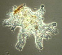 |
The first two photographs are of
an unknown species of amoeba and were taken with phase
contrast at a magnification of 250x. The pseudopodia,
which tend to branch out in all directions searching for
that tasty morsel are one of the main features of this
type of amoeba. Detail otherwise missed can clearly be
seen in the two photographs. The cytoplasm of this
species is differentiated into two distinctive zones, the
inner fluid part, which is called the endoplasm and the
somewhat clearer outer region which is in the form of a
gel. |
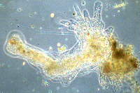 |
 This photograph is of the same species but taken
with a darkground condenser in place. Many crystals and
inclusions within the cytoplasm are clearly visible. Direct rays
from the light source are prevented from entering the front lens
of the objective by a stop which is situated in the substage
condenser. Bacteria, flagella and cilia benefit greatly from this
type of illumination.
This photograph is of the same species but taken
with a darkground condenser in place. Many crystals and
inclusions within the cytoplasm are clearly visible. Direct rays
from the light source are prevented from entering the front lens
of the objective by a stop which is situated in the substage
condenser. Bacteria, flagella and cilia benefit greatly from this
type of illumination.
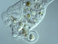 These last two photographs were taken with a
Nomarski system which gives the illusion of a 3D effect and
improves upon the resolution of what can be seen when looking at
algae and protozoa, due to the optical sectioning of the specimen
by the illumination. The light
These last two photographs were taken with a
Nomarski system which gives the illusion of a 3D effect and
improves upon the resolution of what can be seen when looking at
algae and protozoa, due to the optical sectioning of the specimen
by the illumination. The light 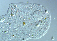 is first polarised and then passed
through a prism which splits the light up into two beams and then
rotates them so they are at right angles to each other, the light
then travels through the specimen and up to the objective where
the beams recombine and interfere with each other. This method is
particularly expensive to buy but if the opportunity to use such
a piece of equipment occurs grab it with both hands The first
photograph show a food vacuole with the remains of some
micro-organism still in the process of being digested, while the
final shot is that of the very granulated nucleus which stands
out clearly. Notice the lack of any colour variations when using
Nomarski.
is first polarised and then passed
through a prism which splits the light up into two beams and then
rotates them so they are at right angles to each other, the light
then travels through the specimen and up to the objective where
the beams recombine and interfere with each other. This method is
particularly expensive to buy but if the opportunity to use such
a piece of equipment occurs grab it with both hands The first
photograph show a food vacuole with the remains of some
micro-organism still in the process of being digested, while the
final shot is that of the very granulated nucleus which stands
out clearly. Notice the lack of any colour variations when using
Nomarski.
The types of
microscopes that I use are a Leitz Orthoplan large field
microscope fitted with an Orthomat fully automatic camera, which
has a built in zoom optical attachment. The exposure range is
almost unlimited with this camera. The lenses are either planapo
or plan fluorite. A Carl Zeiss microscope is also used with very
similar specifications.
Comments
on the article to the author Steve
Durr
© Microscopy UK
or their contributors.
Please report any
Web problems or offer general comments to the Micscape Editor,
via the contact on current Micscape Index.
Micscape is the
on-line monthly magazine of the Microscopy UK web
site at Microscopy-UK
WIDTH=1
© Onview.net Ltd, Microscopy-UK, and all contributors 1995 onwards. All rights
reserved. Main site is at www.microscopy-uk.org.uk with full mirror at www.microscopy-uk.net.