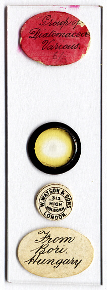
This is an historical slide by W. Watson&Sons, London. Without a magnifying device the faint ash-grey spot in the center of the black lacquer ring is at best hardly visible and might be interpreted as some water staining or a tiny trace of fat. But what a difference when the same slide is viewed under a microscope - have a look yourself at the images below! I think that the visual impression of this beautiful slide needs little or no comment. So please, take some time, enjoy the slide like a real enthusiast, look at the images thoroughly and keep in mind the patient work that has been done by the mounter before you pass to the technical details below. |
|
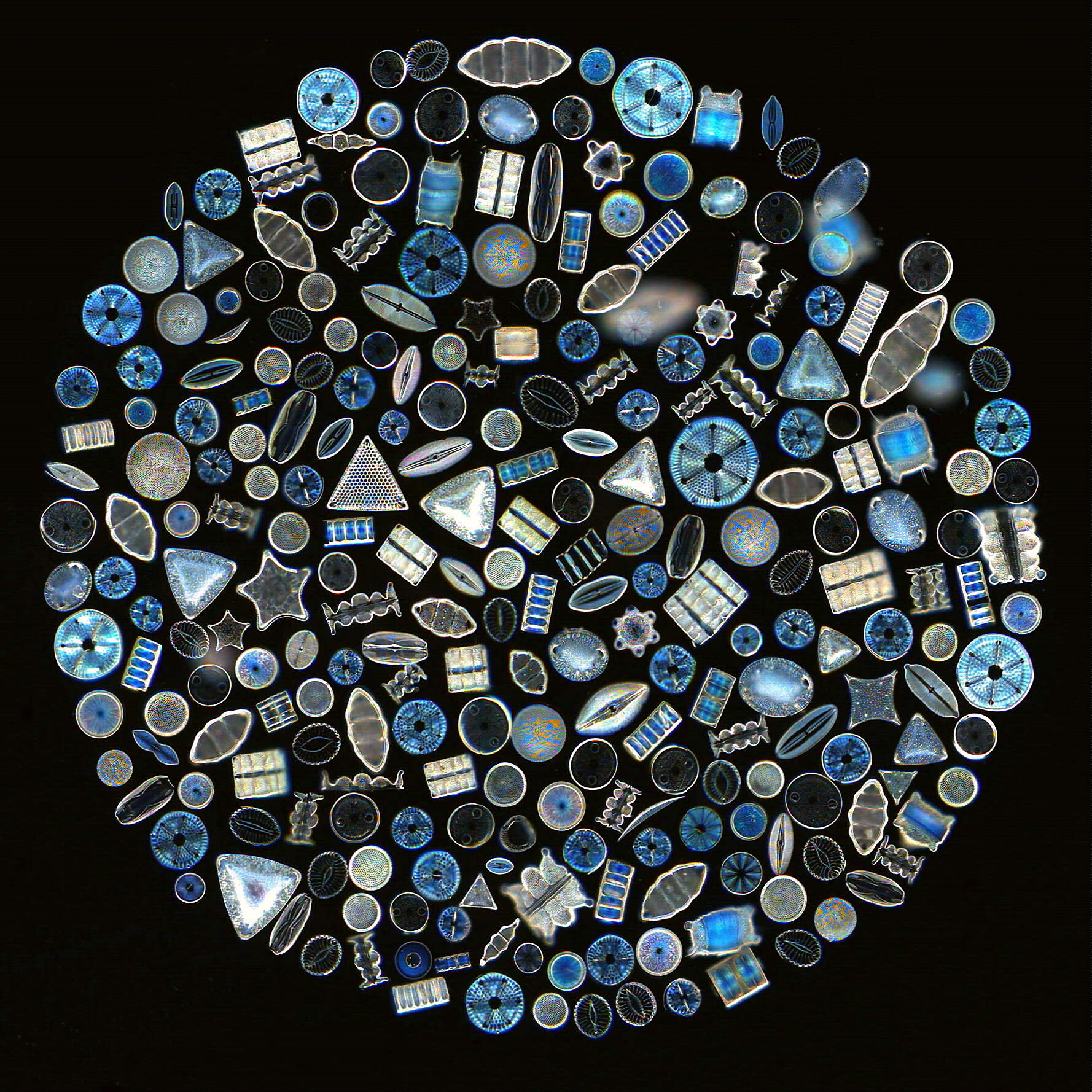
Click on the image to view a 520kB enlargement showing the magnificent detail.
The slide size is 75 mm x 25.5 mm. The diatom circle has a diameter of 2 mm.
The overall view of the slide has been made with a Plustek 600 dpi flatbed scanner which is very affordable (its cost is equivalent to about 60 ordinary hamburgers in Germany). The image processing software Micrografx Picture Publisher 8.0 had been provided on the flatbed scanner installation CD.
The photomicrograph has been taken with a 10x / N.A. 0.30 microscope objective. It is the result of a combination of about 30 individual 640x480 webcam CCD pictures. The camera used was a 'Compro PS39' web camera. The darkfield has been produced by means of a black 1 cm diameter paper disk in the filter tray of the microscope (the exact diameter depends on your equipment, see this Micscape article).
Some of the diatom shells are very much out of focus, probably because they had already lost contact with the fixing glue at the time when the mounting medium was applied. It would have been no problem to have these areas retouched in the picture by means of the imaging software. Nevertheless I have decided to keep the small deficiencies in order to preserve the authentic character of the original slide as much as possible in the picture.
A discussion of how this type of slide has been made is obviously beyond the scope of this article. But an excellent starting point for those who would like to try themselves can be found in: M.I. Cross / M.J. Cole: Modern Microscopy. Lesson 12. P. 169 - 174. London 1895. By the way: Guinea pig hairs (normal hairs - do not cut the whiskers off the poor creatures!) are a very good tool for this work, much better than the finest metal needles.
Comments to the author Martin Mach welcome.