
By Ian Walker UK.
The following pictures were taken on a
Nikon Coolpix 4500 digicam together with a Zeiss microscope and objectives
plus Leitz Periplan 6.3X, 10X eyepieces.
*Note that all of the pictures have been
post edited with Photoshop Elements to improve contrast and definition
and the magnification shown for each image refers to those seen through
the eyepiece not taking into account the final image size*
The slides in order of the images shown
in the article.
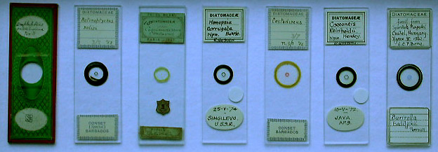
Central image at 63X, outer images
at 250X.
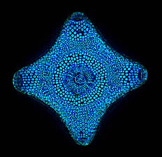
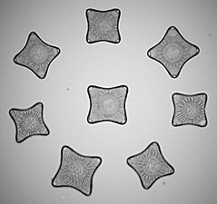
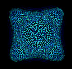
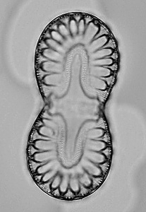
The central image shows a slide by an unknown
mounter, not dated, of eight nicely arranged Amphitetras antediluviana
diatoms and on the outside, colour phase contrast images of two of the
diatoms.
Both at
400X.
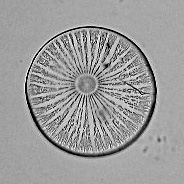
Actinoptychus solisi
from Conset, Barbados, mounted in 1953 by an unknown mounter, on the right,
the same image in phase contrast.
Both at 250X.
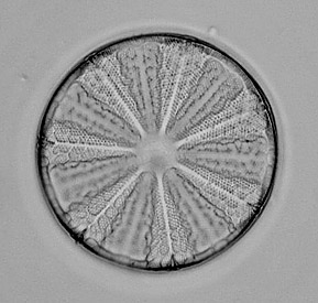
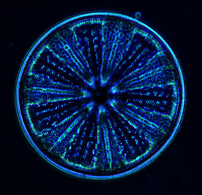
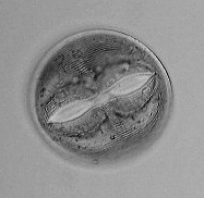
An excellent clear slide from A. C. Cole,
not dated, of fossil Coscinodiscus coucavus diatoms fom Bolivia,
on the right, the same image in phase contrast.
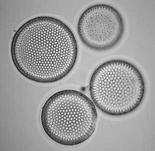
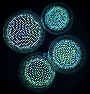
On the left, Monopsia corrugata
from Singilevo, Russia, mounted by R. Gosden in 1974, 250X.
In the middle Cestodiscus from Conset,
Barbados, unknown mounter, mounted in 1952, 250X.
On the right, Cocconeis reinholdii
Hendey from Java AP.9, mounted by R. Gosden in 1972, 400X.
250X
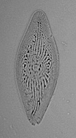
Fossil diatom, Surirella baldjikii
Norman from Castel, Hungary mounted by E. C. P. Bone in 1962.
Comments to the author, Ian
Walker, are welcomed.
the end.
© Microscopy UK or their
contributors.
Published in the April
2003 edition of Micscape Magazine.
Please report any Web problems
or offer general comments to the
Micscape
Editor.
Micscape is the on-line monthly
magazine of the Microscopy UK website at Microscopy-UK
WIDTH=1
© Onview.net Ltd, Microscopy-UK, and all contributors 1995 onwards. All rights
reserved. Main site is at www.microscopy-uk.org.uk with full mirror at www.microscopy-uk.net.