|
|
A Close-up View of the Wildflower "Rose Moss" (Portulaca pilosa) by Brian Johnston (Canada) |
|
|
A Close-up View of the Wildflower "Rose Moss" (Portulaca pilosa) by Brian Johnston (Canada) |
For as long as I can remember, Rose Moss has bloomed along the same section of a driveway near my home, in the quarter inch crack between asphalt and curb. This colourful plant was introduced from South America (the dry plains of Brazil, Uruguay and Argentina), and seems to grow under the most extreme conditions of heat and lack of moisture. It is similar to the popular annual Garden Portulaca which has much larger and more complex blooms.
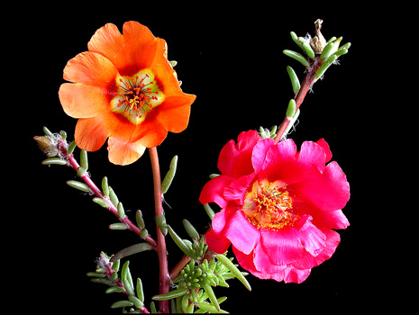
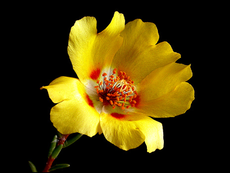
The image above shows two Rose Moss plants. Notice that they have the look of a succulent, with fleshy, torpedo shaped leaves, and perfectly round, reddish stems. The brilliant colours of the blooms range from red through pink, orange and sometimes, yellow. The flowers, which are from two to three centimetres in diameter, open during full sun, and close on cloudy days and at night. The plants grow from five to twenty centimetres high, but the ones that I have observed on the driveway seem to have a maximum height of about ten centimetres. They trail along the ground for most of their length.
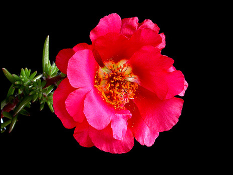
Many flowers, like the one on the left above, have only one ring of petals. The image below shows a bright red bloom with two rings.
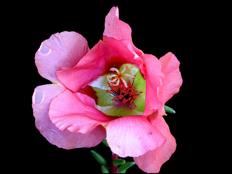
The partially opened pink bloom below also has two rings of petals. Notice that the centre of Rose Moss flowers has a light green, yellow or white colouration.
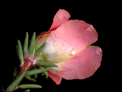
A side view of the flower above reveals the ring of green sepals beneath the bloom.
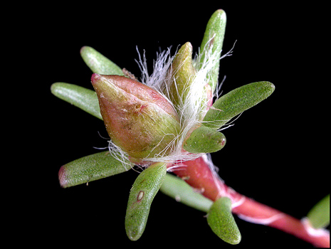
In the image below, the strange clusters of cylindrical leaves are visible. The tufts of whitish hairs are characteristic of Rose Moss.
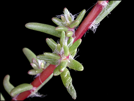
At the end of a stem, a bud about to bloom is also encircled by these fine white hairs.
The following image shows a particularly beautiful flower with a dark red spot near the base of each petal.
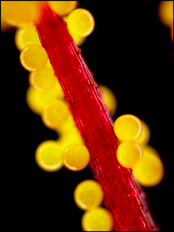
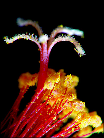
A closer view reveals many stamens, consisting of anthers covered by orange pollen, and supported by dark red filaments. (The anther is the male pollen producing organ.) As well, the pale yellow stigma is visible. (The stigma is the female organ on which pollen lands.)
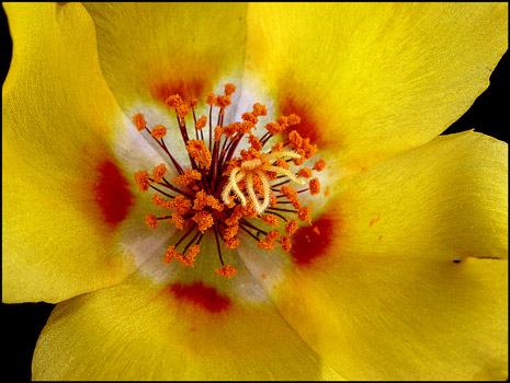
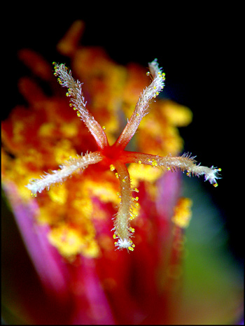
At a higher magnification, the stigma can be seen to be covered with fine hairs.
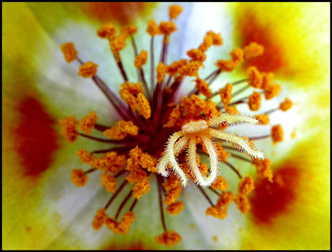
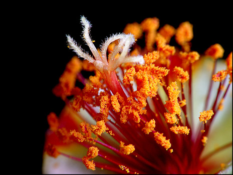
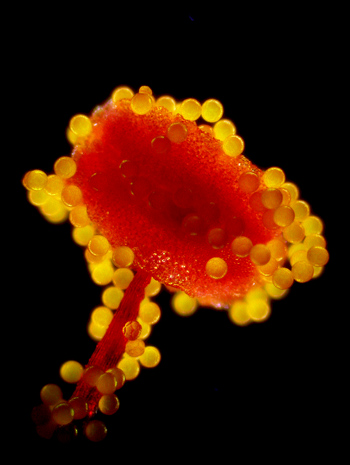
In order to get in close to the stamens and pistil, I removed the petals from a flower. Notice the red circular opening in the end of the style where it spreads out to form the six radial arms of the stigma.
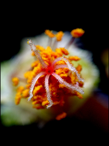
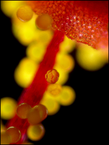
A side view reveals the orange pollen grains coating each anther.
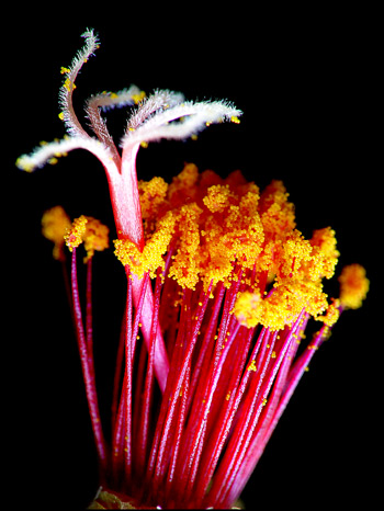
Another side view shows the many shiny red filaments supporting the flower's anthers. The sturdy red style topped by the stigma can be seen protruding above the array of pollen covered anthers. Notice also, the pollen grains which have become attached to the fine hairs of the stigma.
Under the microscope, using dark-ground illumination, a single pollen covered filament can be seen to have spiked ridges. The pollen of the Rose Moss plant is perfectly spherical and appears to have a rough, rather than smooth surface.
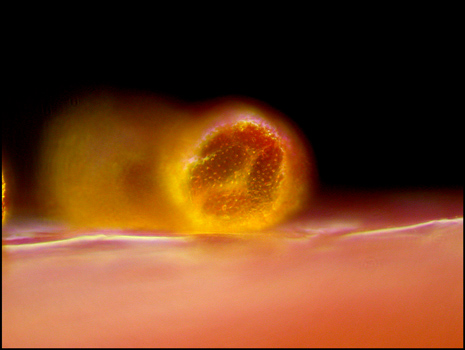
At a lower magnification, an anther sits atop the filament.
A closer view shows a small interloper pollen grain clinging to the filament at the centre of the field of view. Its structure is interesting, as it has a pentagon shape on its front surface. At the top of the image, notice the very rough surface of the anther.
An extreme close-up of the interloper pollen grain reveals more detail.
Although most of the flowers that I observed had large numbers of pollen on their anthers, they had very few grains on the stigma. This leads me to suppose that the pollen of Rose Moss is too heavy to be transported by wind currents, and must be carried by insects. The colourful flowers certainly attract many bees and butterflies.
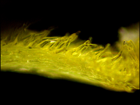
Magnification of the fine hairs on the surface of one of the radial arms of a stigma shows that they are really translucent stalk-like protuberances.
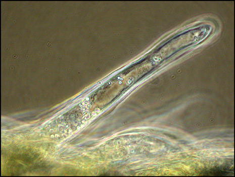
The image below shows one of these protuberances illuminated by phase contrast.
Portulaca grandiflora, a plant very similar to Rose Moss, has much larger flowers with more rings of petals, and more complex patterns. It is available commercially as an ornamental cultivar. Often, the seeds of this plant escape from gardens and the resulting generations may slowly regress to a simpler form. Since I can't find any other Rose Moss plants in the vicinity, I wonder whether this has happened to produce the plants photographed in this article. The closest Portulaca grandiflora plants are about half a kilometre from the driveway.
No matter how they came to be in their present location, one must admire these little plants for their tenacity in the face of extreme conditions, including winters where the temperature dips to much below zero Celsius for prolonged periods. Based on past experience, I fully expect these little marvels to bloom again next August as they have done so many times before!
Photographic Equipment
The low magnification photographs in the article were taken using a Nikon Coolpix 4500 with natural light and the Nikon Cool light SL-1. Higher magnification images were taken using a Sony CyberShot DSC-F 717 equipped with an accessory achromat which screws into the 58 mm filter threads of the camera lens. (This produces a magnification of from 7X to 9X for a 4x6 inch image.) Still higher magnifications were obtained by using a macro coupler (which has two male threads) to attach a reversed 50 mm focal length f 1.4 Olympus SLR lens to the F 717. (The magnification here is about 13X for a 4x6 inch image.) The photomicrographs were taken with a Leitz SM-Pol microscope (using dark ground and phase contrast condensers), and the Coolpix 4500.
All comments to the author Brian Johnston are welcomed.
Please report any Web problems or offer general comments to the Micscape Editor.
Micscape is the on-line monthly magazine of the Microscopy
UK web
site at Microscopy-UK