|

|
A
Close-up View of Wild Cherry Blossoms
(Genus Prunus)
by Brian Johnston (Canada)
|
Loveliest of trees, the
cherry now
Is hung with bloom along the bough,
And stands about the woodland ride
Wearing white for Eastertide.
------
A. E. Housman
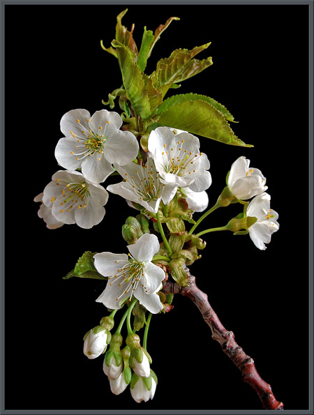
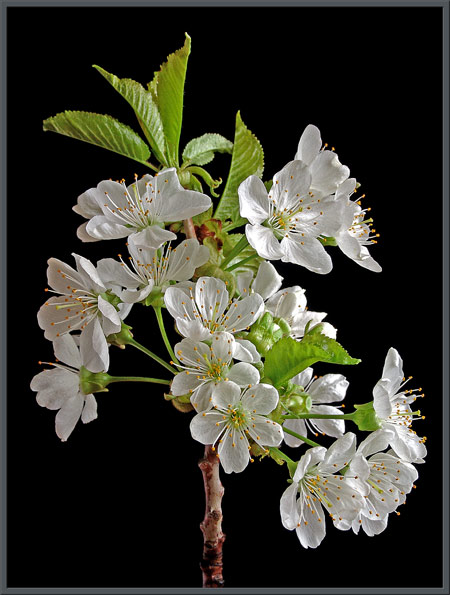
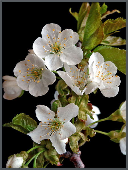
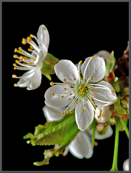
A wild cherry tree grows next to
the bank of a small stream near my
home. For much of the year it looks unkempt, wild and straggly.
In early spring however, a transformation takes place, and the tree is
literally smothered in beautiful white blossoms like those shown above.
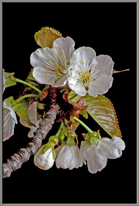
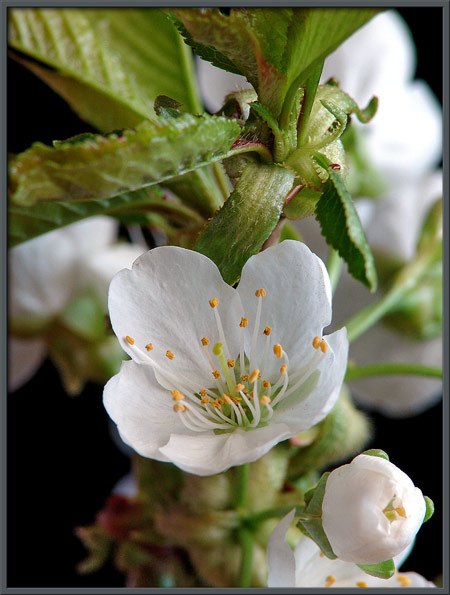
A few days earlier, the tip of the
same branch had buds and partially opened flowers. I was
surprised to find that a branch of flowers, when cut and placed in
water, continues to grow for more than a week. This meant that I
didnt have to keep cutting new samples from the tree to use as
photographic subjects.
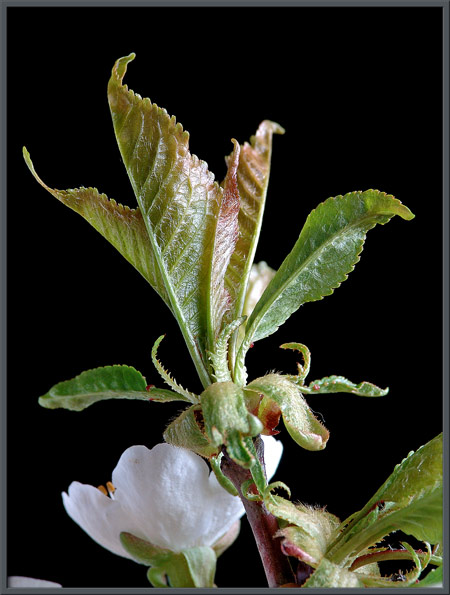
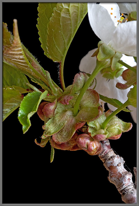
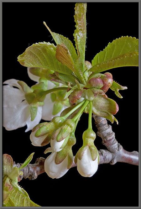
Notice below, the early leaves that
have not opened fully. The pinkish hue slowly fades to light
green as time passes. Strangely, the leaves emerge from a ring of
green sepals (modified leaves).
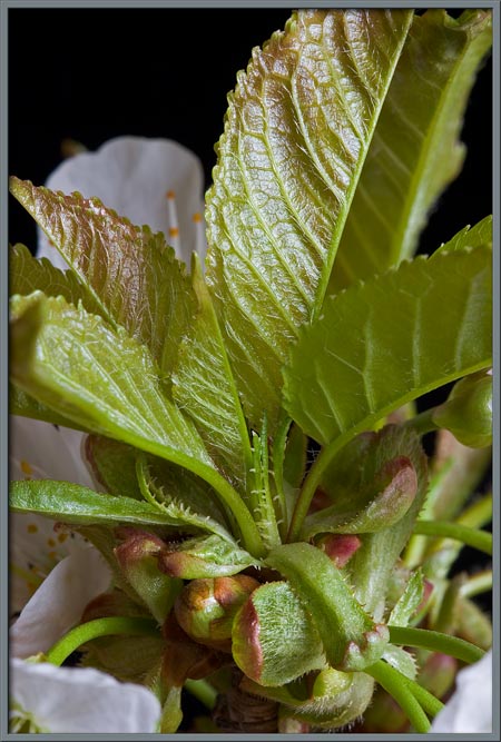
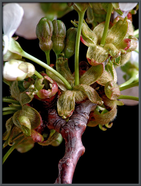
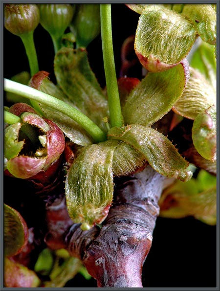
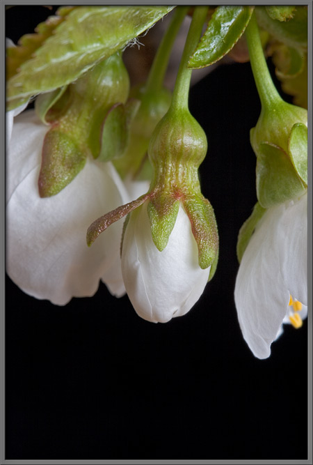
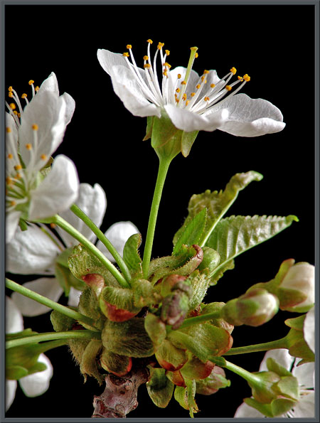
The images below show that the
stems of the buds and blossoms also grow from a similar ring of sepals
that is right at the bark level. From one to four or five stems
can emerge from a single sepal ring. Twigs are reddish-brown in
colour, but tend to become grayer later in the season.
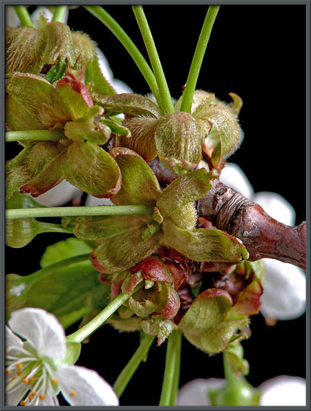
Immature sepals are quite curved,
and have many sharp, pointed serrations along their edges.
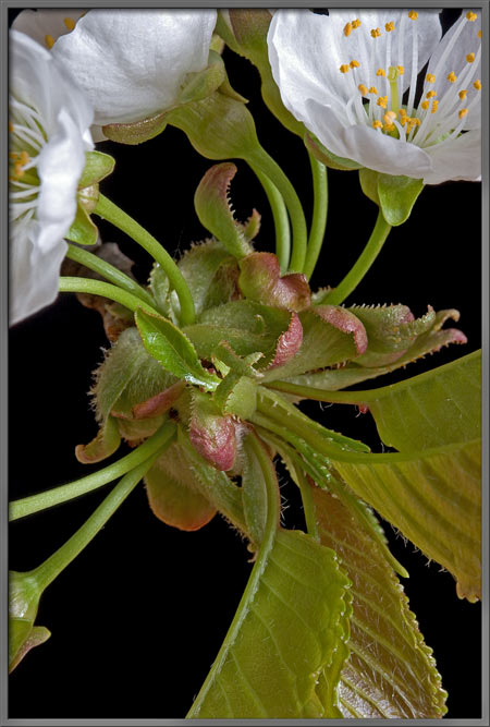
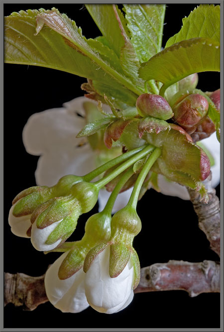
The developing buds are completely
white in colour, and have a ring of five light green sepals clasping
them. Between the bud and stem is a bulbous growth-the ovary (the organ that develops into
the fruit). In several of the images, you may notice a hint of
the bright yellow anthers within the bud.
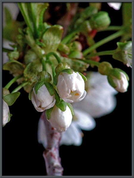
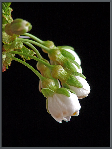
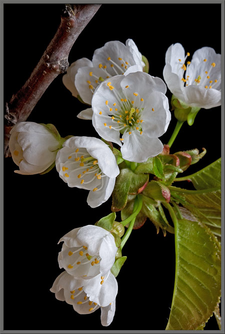
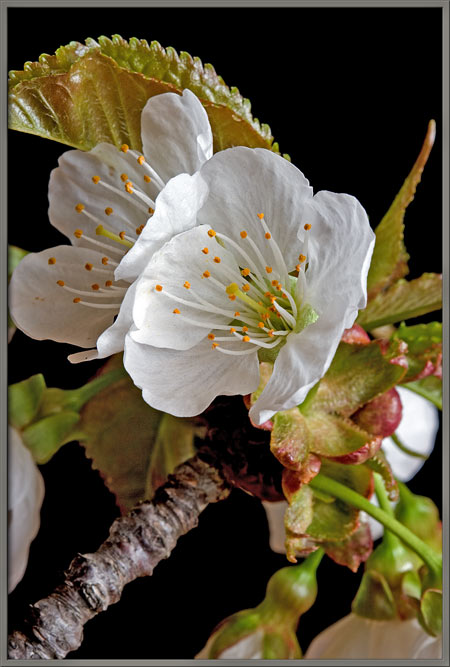
Flowers in various stages of
opening are shown in the image that follows.
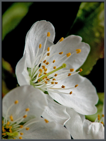
Although wild cherry blossoms are
not colourful, they are nevertheless striking!
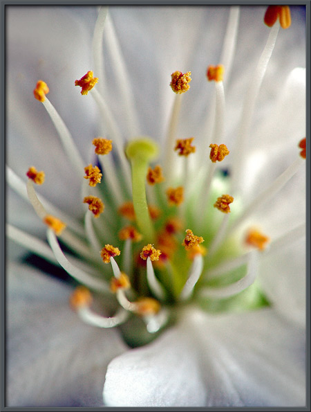
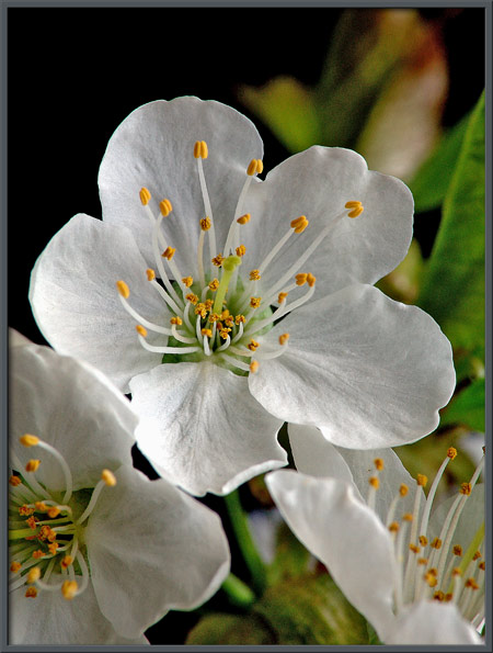
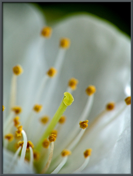
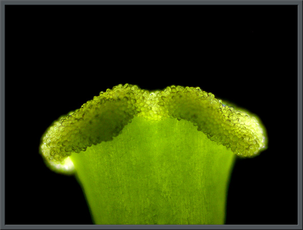
A closer look at a blossom shows
the
details of its reproductive structures. A single pale green pistil, composed of a cylindrical style supporting a bulbous stigma, (the female pollen accepting
organ), is surrounded by many stamens,
composed of thin white filaments
topped by bright yellow anthers,
(the male pollen producing organs).
Up to about twenty stamens surround
the pistil in a given flower. Each anther is usually encrusted
with pollen grains.
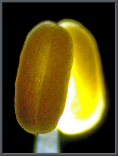
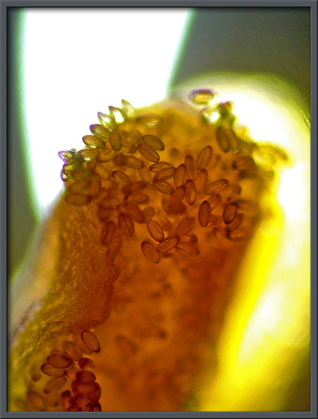
Strangely, there are no pollen
grains on the surface of the anther shown in the photomicrograph on the
left below. Another anther, shown with higher magnification, has
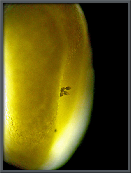
a few ellipsoidal grains stuck to its surface.
This anther has many pollen grains
attached, and the longitudinal groove on each grain is clearly visible.
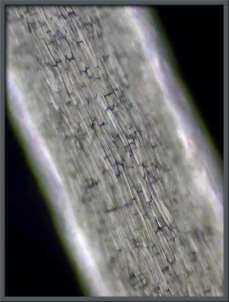
The supporting filament is composed
of many long, tightly packed rectangular cells.
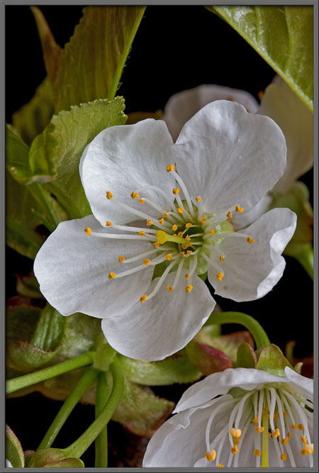
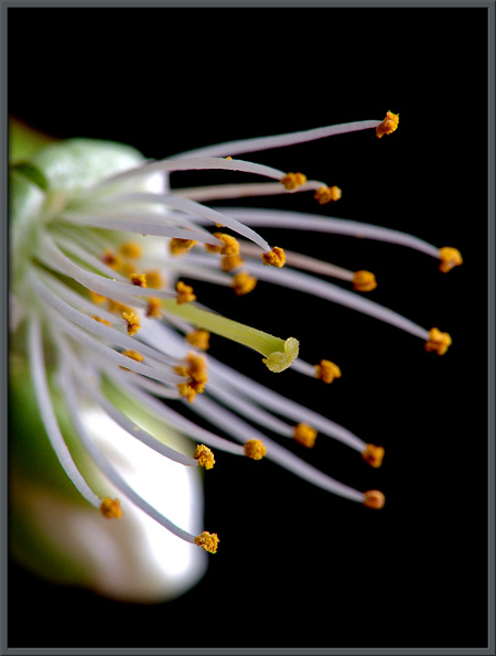
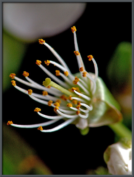
The single pale green pistil in
each flower is about the same length as the anthers.
With the flowers petals removed,
it is easier to see the distinctively shaped stigma at the tip of the
style.
The stigma is almost perfectly
heart-shaped. A photomicrographic side view of the stigma shows
the many blunt protuberances that capture pollen provided by insect
visitors - mainly honeybees. A wild cherry flower is self-sterile, and since the
flowers own pollen cannot fertilize it, insects are essential.
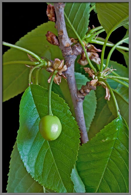
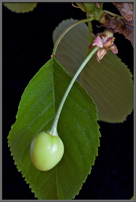
Once a flower has been successfully
fertilized by an insect, the ovary swells, and a transformation begins.
Several weeks later, the fruit has
become a recognizable cherry.
Photographic Equipment
About a third of the photographs in
the article were taken with an eight megapixel Canon 20D DSLR and Canon
EF 100 mm f 2.8 Macro lens. An eight megapixel Sony CyberShot
DSC-F 828 equipped with achromatic close-up lenses (Canon 250D, Nikon
6T, Sony VCL-M3358, and shorter focal length achromat used singly, or
in combination), was used to take the majority of the images. The
lenses screw into the 58 mm filter threads of the camera lens. Still
higher magnifications were obtained by using a macro coupler (which has
two male threads) to attach a reversed
50 mm focal length f 1.4 Olympus SLR lens to the F 828. The
photomicrographs were taken with a Leitz SM-Pol microscope (using
dark-ground condenser), and the Coolpix 4500.
Reference
The following reference has been
found to be valuable in the identification of trees, and it is also a
good source of information about them.
- Little, Elbert L. National
Audubon Society Field Guide to North American Trees - Eastern Region.
2004. Alfred A. Knopf, Inc. (Chanticleer Press, Inc. New York)
©
Microscopy UK or their contributors.
Published in the April
2006 edition of Micscape.
Please report any Web problems or
offer general comments to the Micscape
Editor.
Micscape is the on-line monthly magazine
of the Microscopy UK web
site at Microscopy-UK
©
Onview.net Ltd, Microscopy-UK, and all contributors 1995 onwards. All
rights reserved. Main site is at www.microscopy-uk.org.uk
with full mirror at www.microscopy-uk.net .