|
One of "Nature’s Little Secrets" An Unknown (to me) by Richard L. Howey, Wyoming, USA |
As Heraclitus said: “Nature loves to hide.” For the naturalist, it is almost always a contest between oneself and the elusive and secretive character of nature. Sometimes I think that Sir Francis Bacon’s observation that one has to put Nature on the rack and torture answers out of her, was not so far off the mark. Over the last 2,500 years, humans have stumbled and bumbled around trying to figure out how natural phenomena work. Amazingly, given the human predisposition to resort to mythical, mystical, and theological accounts for things they can’t yet explain, some fairly significant progress in the way of rational, empirical, and scientific explanation has been made.
A major complication is that even after a specific secret has been extracted, revealed, and published, it often remains very difficult to access. These days, scientific journals are frequently so highly technical that only the specialists can understand them and, furthermore, they are usually so expensive that only research institutes, universities, and special libraries can afford them. More and more, scientific papers are being published electronically in online journals, but for many of these one has to pay for a subscription. Furthermore, there is the enormous problem of getting back issues of thousands of scientific publications online in dozens of languages and, some of these journals have histories that date back over a century and a half. The complexity of the task is enormous and the cost would be staggering. If we undertook projects like this, we wouldn’t be able to afford to have wars and that simply wouldn’t do.
So, for now, we have to rely on the expertise and good will of other fellow enthusiasts, be they amateur or professional, and that’s what I’m relying on in this case. A few weeks ago, I was looking at some preserved specimens of the tunicate Molgula manhattensis and on two of them, I noticed some odd little things attached to the outer sandy surface of the Molgula. There must have been a hundred or more of them. Here’s one example.
At first I thought it might be another species of tunicate, since it clearly had an aperture that might be a siphon.

Look as hard as I might, I couldn’t find a second siphon and a tunicate with only one siphon would be like a one-wheeled bicycle–and, yes, I’ve hear of unicycles, which are by definition not bicycles! In nature, there are, of course, always exceptions: mutants, monster formations, genetic aberrations, and all sorts of other delightful mischief, so I decided to try a small test. Usually the outer casing, test, or tunic is composed largely of cellulose compounds. On previous occasions, when I was trying to isolate calcareous spicules in sessile (attached) tunicates (also known as ascidians), I took thin sections of the tunic and subjected them to treatment with sodium hypochlorite or potassium hydroxide. [Caution: These are both highly caustic substances.] The tunic sections were highly resistant to these caustic agents and so were useless for that purpose. A very wide range of animal tissues are attacked and dissolved by these caustics. So, I took one of my strange unknowns, put it on a slide with a drop of sodium hypochlorite and it immediately began to dissolve. Conclusion: It almost certainly is not a tunicate. I took images of a couple of other specimens from different angles using different sorts of illumination and I’ll show you two of them here.
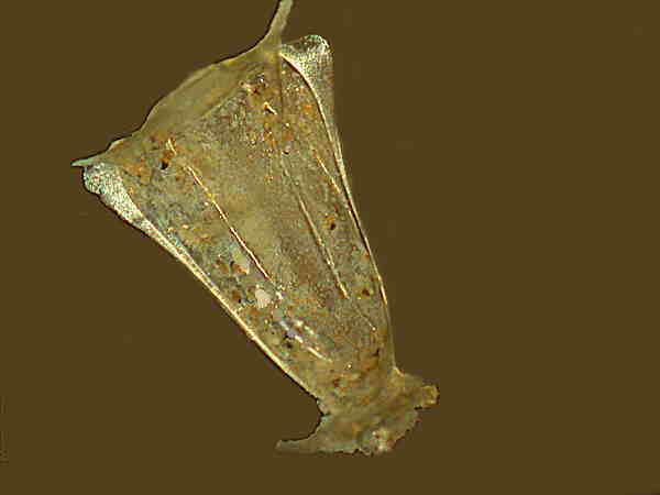
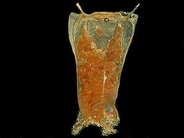
This is a side view taken with brightfield illumination with the bottom end on the right. The irregularly shaped structure on the bottom end is a holdfast or “anchor” by means of which it attached itself to the Molgula.
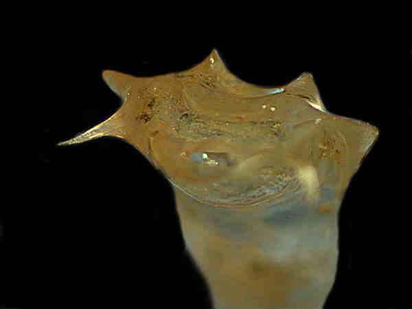
This image is a side view of the top with oblique illumination. You can see the pore or orifice and the horn-like structures around the top, two of which are considerably longer as we’ll see in the next two images. All of these specimens are virtually the same size and are about 3 mm. in length.
I had removed almost all of these unknown thingies from the bodies of the Molgula and placed them in a separate micro-jar of 70% alcohol. When I next examined the jar, I noticed that some of the specimens were floating at or near the surface while some others were on the bottom of the jar and looked darker. I extracted 3 specimens from the bottom and it was immediately obvious that they, unlike the others I had looked at, were packed with lots of tiny round things. Again I’ll give you 2 views.
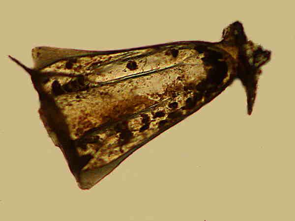
The first image is brightfield and shows the specimen upright with the two longer “horns” clearly visible and the holdfast at the bottom. They tend to cluster around the siphons of the tunicate which suggests that they get benefit from the currents which the siphons create.
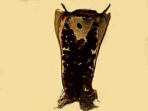
Here is the same specimen with oblique illumination and a computer-generated black background for contrast. Immediately I was curious about all of these pinkish-peach colored “spheres” inside. This prompted a bit of micro-surgery to extract some of the “spheres” to be examined at higher magnification with a compound microscope. So far, all of my observations and imaging had been done with my trusty old Olympus SZ-III stereo-zoom microscope. Here are the results I got with the compound microscope.
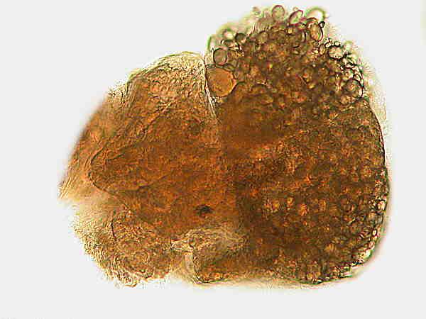
The first glance suggested to me that this was not simply an egg, but an egg that had begun to develop, perhaps an early larval stage.
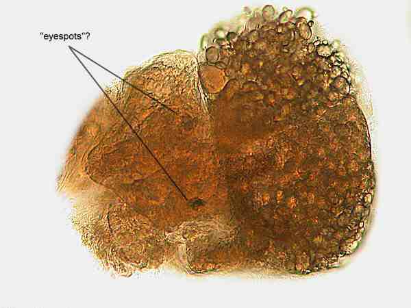
Something that drew my attention was the two dark spots near the center of the mass. I suspect that these may be “eyespots” and it may well be that as these larvae develop, the eyespots become more complex.
So, my final uneducated guess is that these long tubular structures are egg cases and that they contain the larvae of some kind of small mollusk, perhaps a gastropod. I don’t know quite why, but the general impression I gained was that this creature was snail-like in its larval appearance. I may very well be wrong and I am appealing to Micscape readers to help me identify this intriguing organism. The first one to correctly identify it will get a mention in my next article and a free subscription to my newsletter, if I ever get around to starting one.
Finally, I suspect that many of you encounter, from time to time, strange critters which you can’t identify. I strongly encourage you to take a few pictures, write up a description, send it to Micscape and give the readers the fun of making some guesses.
All comments to the author Richard Howey are welcomed.
Microscopy
UK Front Page
Micscape
Magazine
Article
Library
Please report any Web problems or offer general comments to the Micscape Editor.
Micscape is the on-line monthly magazine of the Microscopy UK website at Microscopy-UK