|

|
A
Close-up View of the
"Single-Seed Hawthorn"
Crataegus monogyna
by Brian Johnston (Canada)
|
The beautiful Hawthorn, that has now put
on
Its summer luxury of snowy wreaths,
Bending its branches in exuberant
bloom,
While to the light enamour'd gale
it breathes,
Rife as its loveliness, its rare
perfume.
Glory of England's landscape!
Favourite tree
Of bard or lover! It flings far and
free
Its grateful incense.
From the "Forest Minstrel" by William
Howitt
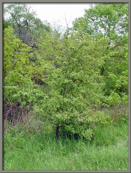
The hawthorn tree photographed for
this article grows in the flood plain of a small river, and is located
about ten metres from the bank. About three metres in height, it
has branches almost to ground level. Strangely, if hawthorns grow
in a forest environment, they tend to shed their lower branches and
look more like normal trees. Since the trees branches possess
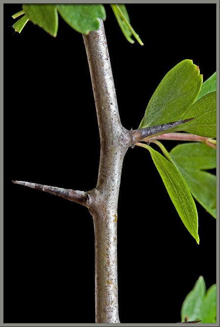
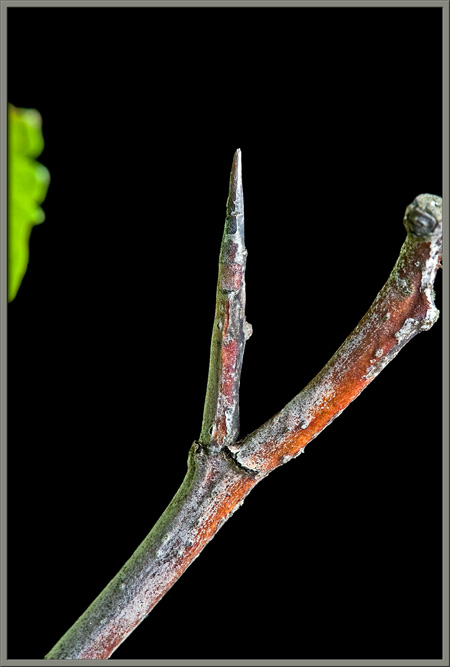
many straight, stout spines, hawthorns have been a popular hedgerow
or boundary tree, used to discourage entry into properties.
Crataegus
monogyna is a member of the Rosaceae
family. The genus name Crataegus
derives from the ancient Greek for strength, and refers to the tough
hardwood of the trunk and branches. (Walking sticks are sometimes
made from hawthorn wood.) Monogyna,
the species name, refers to the fruit of the plant, which has a single (mono) seed (gyna). There are several
common names for this species other than single-seed hawthorn,
including whitethorn, and may.
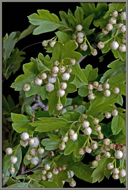
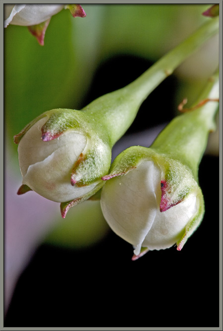
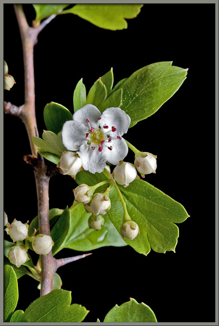
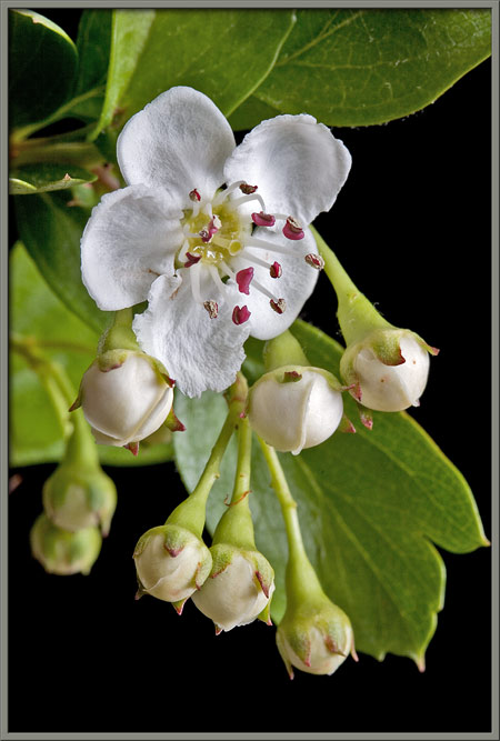
At the beginning of May, the
hawthorn tree is liberally sprinkled with tiny, almost spherical white
buds. The image on the right shows the five, pointed sepals,
(modified leaves), that ring each buds base, and the swellings at the
end of the stalks that indicate the presence of immature ovaries.
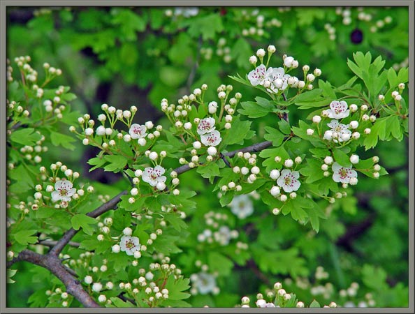
The blooming process occurs over
the relatively short period of seven to ten days. The image that
follows shows this development at an early stage.
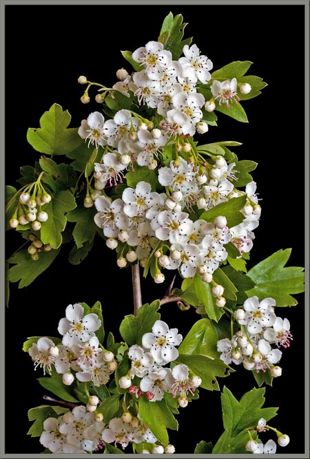
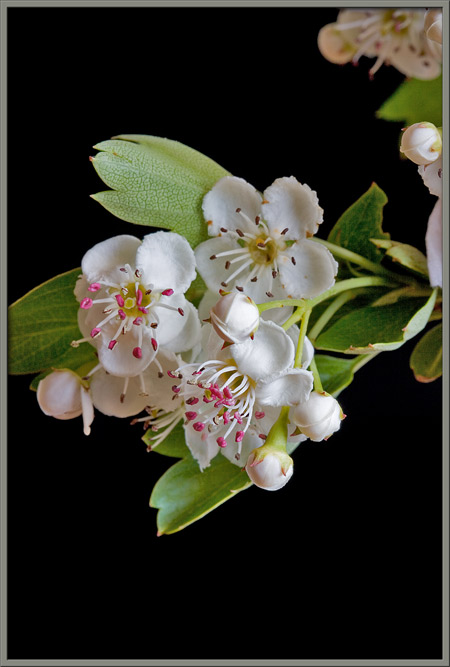
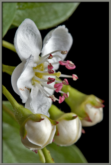
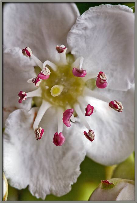
Clusters of white, five-petaled
flowers are so numerous as to almost obscure the trees leaves and
branches. The flowers are often described in the literature as
being scented or heavily scented, which in my opinion, gives the
impression that their smell is pleasant. Perhaps, to some, but I
find the scent to be vaguely fishy and not pleasant at all!
(This may be due to the fact that I bring branches indoors in order to
photograph them, and this concentrates the smell in a smaller volume
than that of the out-of-doors.)
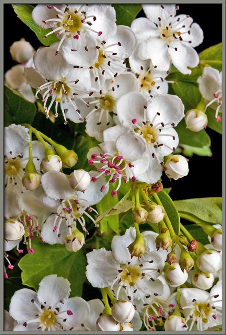
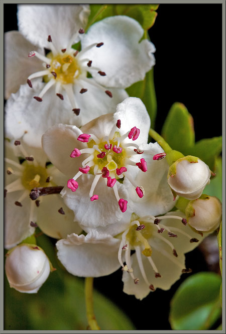
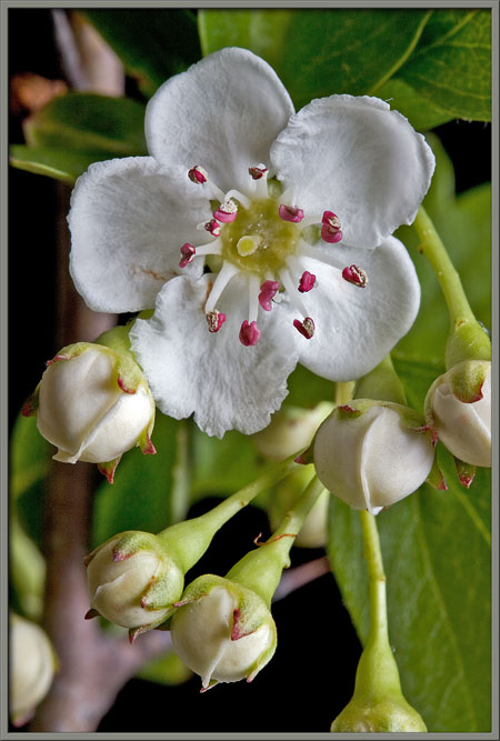
At any given time, a branch has
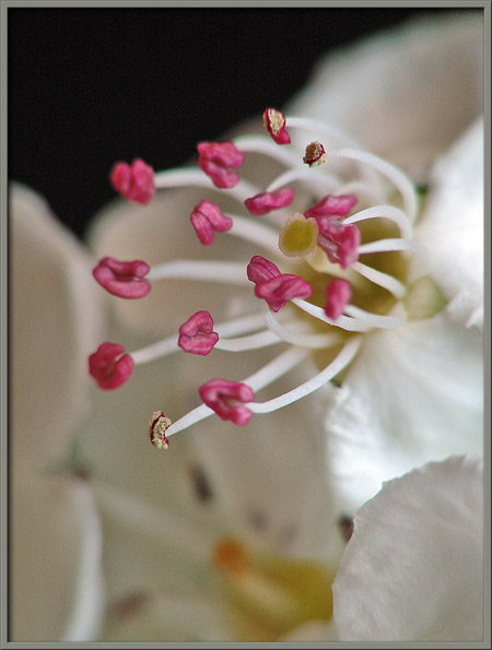
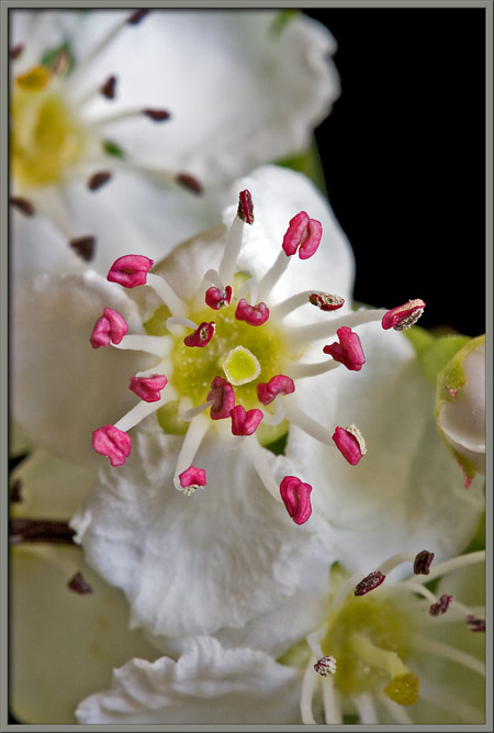
blooms in many developmental stages. Notice below that when a
flower first opens, the anthers are large and a bright red
colour. In less than twenty four hours the anthers appear to
shrink, and take on a dark brown colouration.
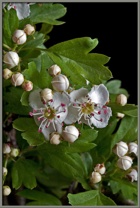
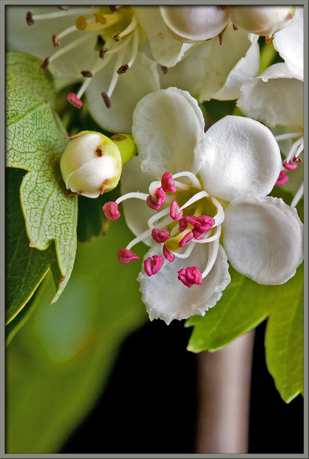
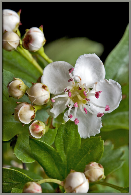
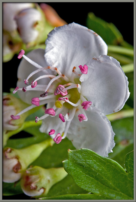
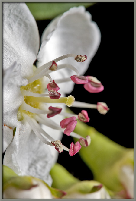
Hawthorn flowers have five,
relatively thick, rounded petals with irregular edges. Notice the
sharp spine in the lower left of the image.
Closer views of spines can be seen
below. The tree is located in an area with no paths, and reaching
it though long grass and vines, that are almost waist height, is a very
difficult task. As if this wasnt enough, every trip to
the tree resulted in blood loss, due to the vicious spines.
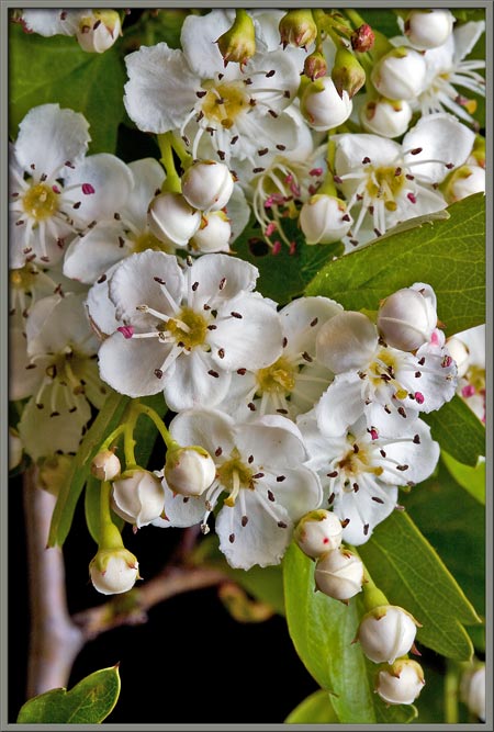
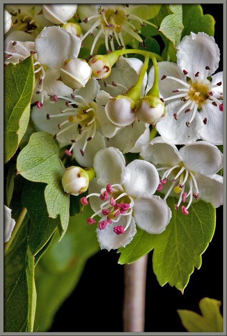
The four images below show typical
flower clusters. Notice the rough edges on petals, and the red
tips of the sepals at the base of each flower.
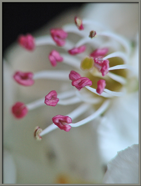
Here are three closer views of
several just opened flowers. Notice in the third image, that
the change in the appearance of anthers has begun to occur.
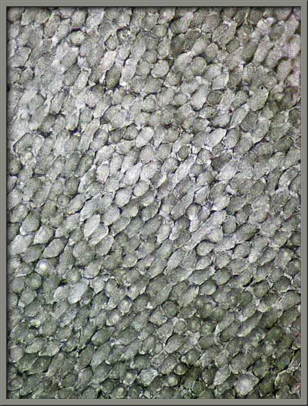
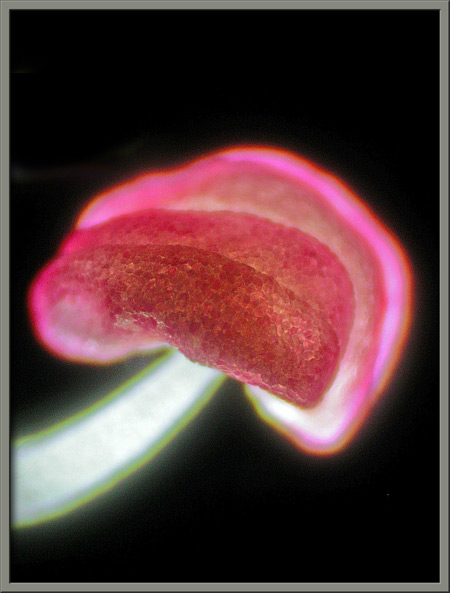
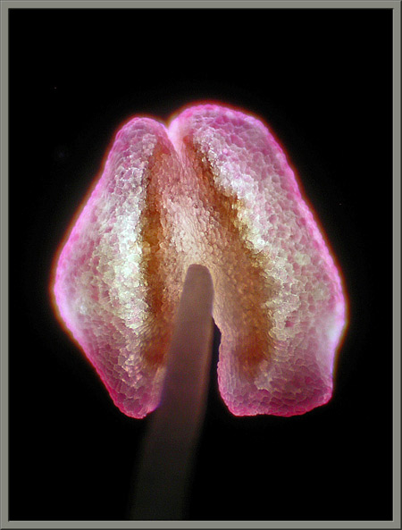
The photomicrograph on the right,
below, shows the cellular structure of a hawthorn flower petal.
A flower has multiple stamens consisting of reddish anthers, (the male pollen producing
structures), supported by white filaments.
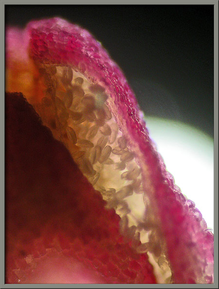
Most anthers appear to have a
pointed horseshoe shape. If you look closely at the images below,
you will see that the red structures do not have pollen on their
surfaces but that the smaller brown structures do! It may be
that what I have referred to as red anthers, were in fact anther caps, that disintegrate to
reveal the actual brown anthers beneath! (I could not, however,
find any supporting evidence for this in the literature.)
Here then, are photomicrographs
showing what I suspect to be an anther cap, and supporting filament.
Looking through a crack in the
anther cap reveals pollen grains with an ellipsoidal shape. (I
am more convinced of my hypothesis!)
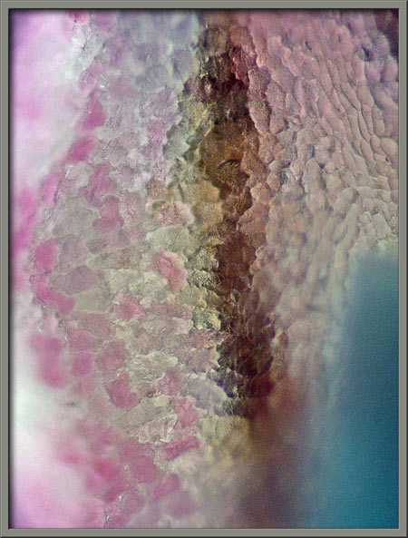
Notice the difference in
pigmentation of cells in various locations on an anther cap.
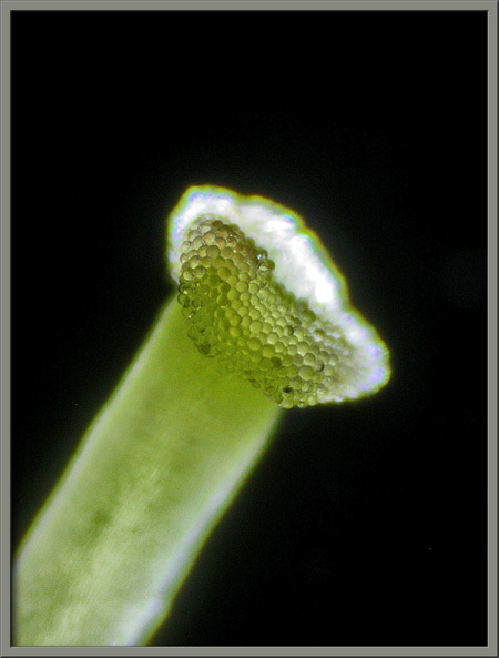
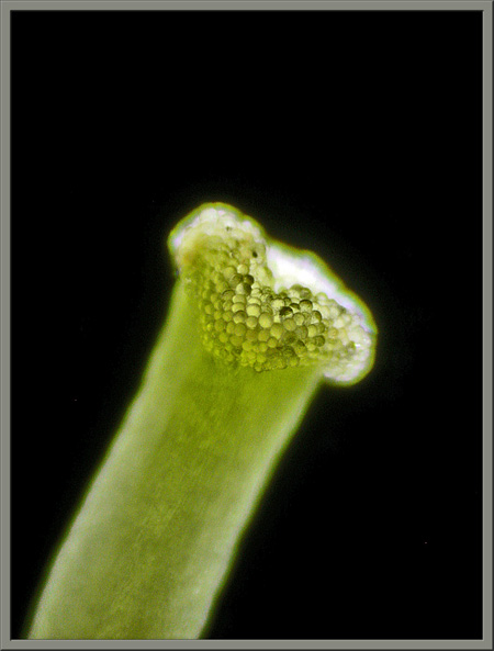
This species of hawthorn has a
single pistil consisting of a
bulbous, light green stigma,
(the female pollen accepting organ), supported by a similarly coloured style. (Further to the earlier
discussion, notice how the pollen seems to be pushing aside the red
anther caps in these images.)
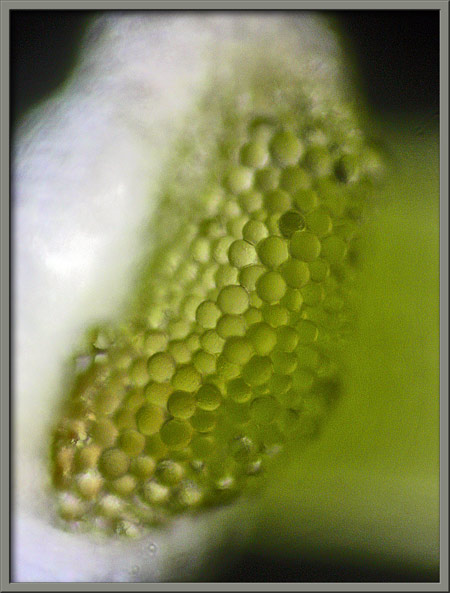
Under the microscope, the stigma
appears to be covered with spherical, glandular protuberances.
These can be seen more clearly in
the higher magnification photomicrograph at left. The cellular
structure of the style is visible in the image at right.
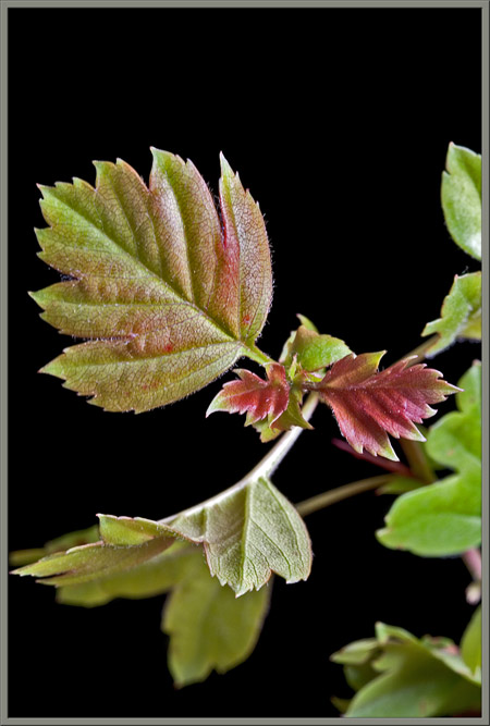
When the trees leaves first
appear, they are bright red, but this colour soon changes to pink, and
then to the final green. The leaves are lobed, and have some
serration.
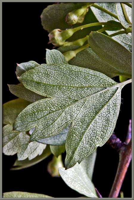
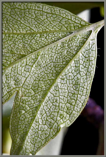
The intricate structure of the
underside of a hawthorn leaf can be seen below.
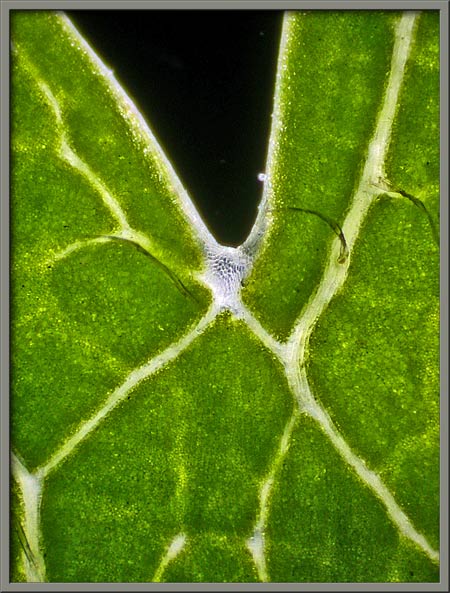
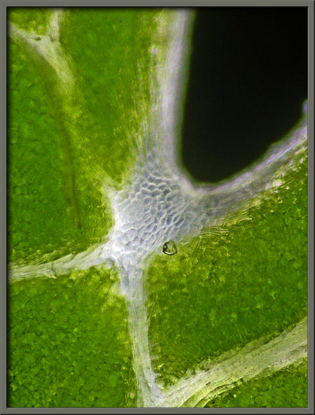
Cells missing green chlorophyll
pigment appear at the intersection of two lobes.
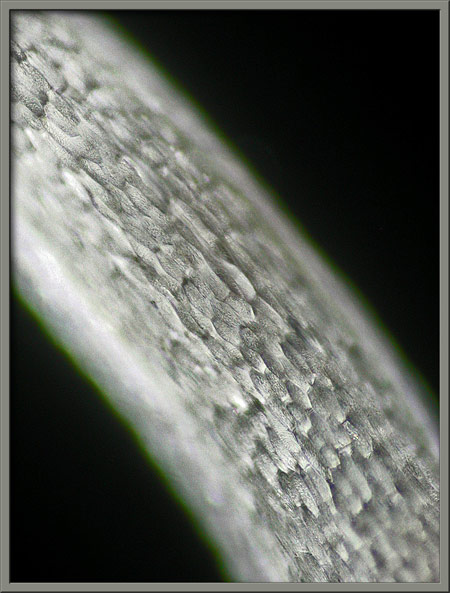
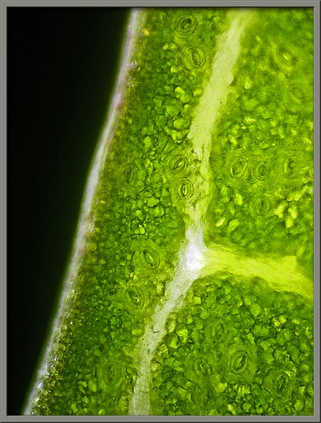
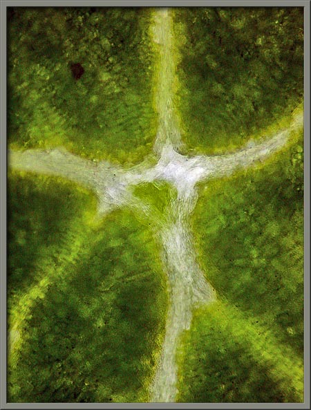
Leaf vein structure can be seen in
the two images that follow. In the image at left, the stoma and guard cells that control the entry
of gas into the underside of a leaf are visible.
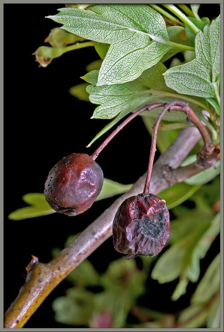
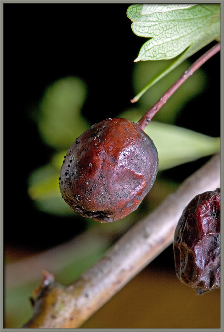
Fertilized flowers produce small
(1.25 cm) red berries called pomes
or haws which contain a single
seed hence the common name single-seed
hawthorn. Fruit eating birds are the primary agents of
seed dispersal in this species. Although germination of the seed
is facilitated by passing through a birds digestive tract, this is not
absolutely necessary for the process to occur!
Several species of hawthorn grow in
my area, but this was the only example of the single-seed variety
that I could find. The difficulty of getting to its location, and
its many blood-letting spines, made this a memorable photo-taking
experience!
Photographic Equipment
Most of the photographs in the
article were taken with an eight megapixel Canon 20D DSLR and Canon EF
100 mm f 2.8 Macro lens. An eight megapixel Sony CyberShot DSC-F
828 equipped with achromatic close-up lenses (Canon 250D, Nikon 6T, and
Sony VCL-M3358 used singly, or in combination), was used to take a few
of the images.
The photomicrographs were taken
with a Leitz SM-Pol microscope (using a dark ground condenser), and the
Coolpix 4500.
References
The following references have been
found to be valuable in the identification of wildflowers, and they are
also a good source of information about them.
- Dickinson, Timothy, et al.
2004. The ROM Field Guide to Wildflowers of Ontario. Royal
Ontario Museum & McClelland and Stewart Ltd, Toronto, Canada.
- Thieret, John W. et al.
National Audubon Society Field Guide to North American Wildflowers -
Eastern Region. 2002. Alfred A. Knopf, Inc. (Chanticleer Press,
Inc. New York)
- Little, Elbert L. National
Audubon Society Field Guide to North American Trees - Eastern Region.
2004. Alfred A. Knopf, Inc. (Chanticleer Press, Inc. New York)
- Kershaw, Linda. 2002. Ontario
Wildflowers. Lone Pine Publishing, Edmonton, Alberta,Canada.
- Royer, France and Dickinson,
Richard. 1999. Weeds of Canada. University of Alberta
Press and Lone Pine Publishing, Edmonton, Alberta, Canada.
- Crockett, Lawrence, J.
2003. A Field Guide to Weeds (Based on Wildly Successful
Plants, 1977) Sterling Publishing Company, Inc. New York,
NY.
- Mathews, Schuyler F.
2003. A Field Guide to Wildflowers (Adapted from Field Book
of American Wildflowers, 1902), Sterling Publishing Company, Inc.
New York, NY.
- Barker, Joan.
2004. The Encyclopedia of North American Wildflowers.
Parragon Publishing, Bath, UK.
©
Microscopy UK or their contributors.
Published in the April
2008 edition of Micscape.
Please report any Web problems or
offer general comments to the Micscape
Editor.
Micscape is the on-line monthly magazine
of the Microscopy UK web
site at Microscopy-UK
©
Onview.net Ltd, Microscopy-UK, and all contributors 1995 onwards. All
rights reserved. Main site is at www.microscopy-uk.org.uk
with full mirror at www.microscopy-uk.net .