John Boyd, microscopist, 1852 - ca. 1930
by Brian Stevenson, Lexington, Kentucky, USA
John Boyd was a relatively well-off Victorian-Edwardian industrialist and amateur microscopist. He was an early member and officer of the Manchester Microscopical Society, the Literary and Philosophical Society of Manchester, and other microscopy clubs. He also amassed what appears to have been a considerable library of microscope slides. Some were probably of his own make, but many were acquired by trading with other microscopists. Slides from his collection, carrying labels with his printed name, pop up for sale fairly often on the internet. Other slides that carry evidence of having been Boyd’s are also frequently seen. This article is intended to help others identify John Boyd’s slides, plus provide both an overview of Boyd’s life and glimpses into amateur microscopist experiences of late 1800s England.
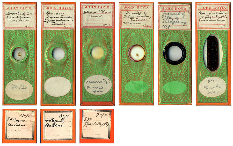
Most of John Boyd’s slides with which I am familiar are wrapped back and sides in plain orange paper, and covered on the front with a green plaid pattern (Figure 1). This plaid pattern appears to have been widely distributed through the late 1800s, as it can also be found on slides from many other sources. However, the pairing of orange with the green plaid seems to be almost unique to Boyd. Many of his slides bear custom-printed labels on their fronts. The handwriting on these labels has occasional inconsistencies, as if Boyd was forcing himself to write in an unfamiliar style and did not always form letters the same ways. For example, in Figure 1, compare the style of “M” in “Mason” on the fourth slide with the same letter in “Moth” on the sixth. On the backs of many Boyd slides are white labels with names, dates and mounting media. This handwriting is probably also Bond’s: notice that the formation of “Balsam” on the first slide of Figure 2 is very similar to that word written on the back labels of slides shown in Figs. 1 and 3.
Figure 1. Examples of papered slides bearing John Boyd’s name plate. All known papered slides of Boyd’s use the same green plaid front paper with orange side and back papering. Below the leftmost three slides are images of labels on their backs, with the apparent sources of these slides: A.C. Rogers, F. Lazenby and E.W. The fourth slide has the handwritten name “Col. Mason” on the front label. The two rightmost slides do not have source information. Note the use of distinctly different ringing cements evident on the fourth and fifth slides. The inconsistencies between the dates on the fronts and those on the backs are a mystery.
Several features of Boyd’s slides indicate that he was not the preparer of most of them. Variously-sized circular and square coverslips were used, with numerous different styles and colors of ringing. This is especially evident in Boyd-labeled slides without wrappers (Figure 2). In addition, many of his slides include notations of other people. Most of those names can be linked to other microscopists or professional slide preparers (discussed in detail at the end of this essay). It appears that Boyd re-wrapped most of his earlier acquisitions in his own style, thereby personalizing and unifying his library.
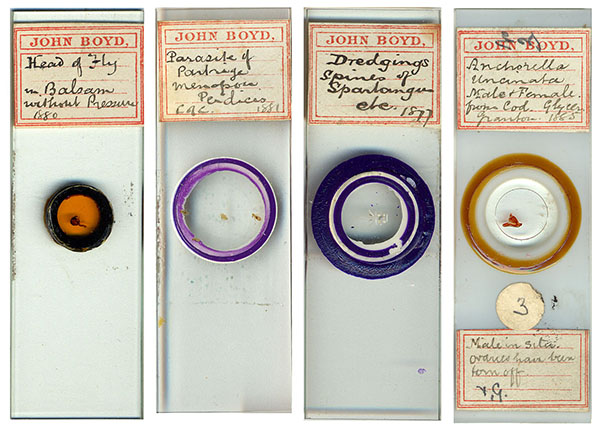
Figure 2. Examples of unpapered slides bearing John Boyd’s name plate. Note that the handwriting of “Balsam” on the leftmost slide is essentially identical to that written on the backs of slides shown in Figures 1 and 3. Notice also that all four examples show different styles of ringing, suggestive of different makers. The rightmost slide is opaque white glass, while the others are transparent.
Other antique slides are known that use the same green plaid with orange wrapping papers, and have the same back-side labels listing sources in the same hand as on the Boyd-labeled slides, but lack John Boyd’s name plate (Figure 3). The handwriting on the oval labels on these slides is rather different from that on the slides bearing John Boyd’s name plate. It may be that Boyd used a more relaxed hand when he wrote out the ovals – there are similarities between many letters on the labels shown in Figs. 1, 2 and 3. Alternatively, these slides may have been labeled by someone else, perhaps a member(s) of one of Boyd’s microscopy clubs. However, I acquired all of the slides shown in Figs. 1 and 3, plus about a dozen more, from the same seller at the same time, who had bought them as part of a larger collection. The coincidence of the identical wrappers, identical labels listing their sources, and the location of all these slides within the same collection strongly support the conclusion that all of the illustrated slides belonged to John Boyd at some point in time.
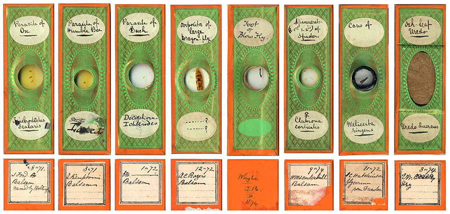
Figure 3. Examples of slides bearing the same green plaid front paper with orange side and back papering used by Boyd, and with similar source information. Extensive similarities between such slides and those illustrated in Figs. 1 and 2 support the conclusion that the above slides were once in John Boyd’s collection. Below each slide is an image of its back, noting the apparent source of the slide. These are, left to right: J. Ford, A. Kempson, E.W., A.C. Rogers, Whyte/J.B., H.M.J. Underhill, J.C. Hutchinson (Hutcheson) and E.W.
John Boyd was born in Manchester, England during the Autumn of 1852. He was the third son of James and Isabella Boyd. James was originally from Scotland, but by the time of John’s birth was a cotton yarn merchant in Manchester. Isabella, and all of their children, were born in Lancashire. The 1861 and 1871 censuses record the family as living in Chorlton upon Medlock, Lancashire. At the time of the 1871 census, 18 year old John was recorded as being a salesman, possibly for his father’s firm.
John evidently had developed a strong interest in microscopy by his early twenties. In 1874, he advertised in Hardwicke’s Science-Gossip for “Well-mounted Diatomaceae, Zoophytes, Palates, Sections, Spicula, &c., for mounted Parasites, showing all the legs.” His address was given as Victoria Park, Manchester. It is interesting that Boyd was offering to provide slides of parasites, since fleas, lice, ticks and the like were a passion of his, as evidenced by his frequent lectures on parasites through the following years. Exchange offers such as this help explain the source notes on the backs of many Boyd slides, they being records of microscopists with whom Boyd swapped slides. The earliest source notes in my collection are from 1870, when Boyd would have been 17 or 18.
In 1875, John Boyd was instrumental in the restoration of the “Leeuwenhoek Microscopical Club” of Manchester. That club was founded in 1867, and consisted of only a small number of members. From A Review of the Work of the Leeuwenhoek Microscopical Club: “Throughout its existence the Club has limited its membership to just that number which its members could conveniently accommodate in each other’s house and seat around a common table; but, generally, at the meetings there has been room for one or two guests. There has never been a period when it could not have enlarged its borders; but a doubling of the number of its members would, in most cases, have proved inconvenient for accomodation in a private house, and would, besides, have consumed too much time in the examination of the suite of objects prepared to illustrate the special study of the evening. It has been found by experience that six or eight persons, with two or three microscopes revolving in order within the circle, make the happy medium for the most effective consideration and discussion of the many points which arise during the demonstration of any practical subject.” The club’s first President was the Rev. John E. Vize, who later achieved fame as a mycologist. The small number of members led to the suspension of the club’s meetings in 1873, after several members resigned due to business obligations, moving away, etc. On Sept. 30, 1875, the club reorganized with a membership of five, including John Boyd. He was elected Secretary, and later served as President from 1890-91. Boyd was also noted in the club archives as having developed a simple rotating table-top platform for moving microscopes from one viewer to another. This club, and probably others of which Boyd was a member, also contributed to his growing collection of microscope slides. The Review notes that “at all these meetings the exchange of slides illustrating the selected subjects of study, or novel modes of mounting, was observed for many years…the cabinets of the members were thus enriched with many valuable slides, which in some cases remain as mementoes of friends passed away.”
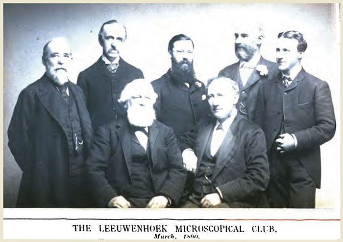
Figure 4. The Leeuwenhoek Microscopical Club, March 1890. The gentlemen are not specifically identified, but include John Boyd, who was 37 years old at the time. The others are Charles Bailey (age 52), John Barrow (age 69), William Blackburn (age 49), John B. Pettigrew (age 36), Mark Stirrup (age 58) and John Tatham (age 45). Pettigrew was single and worked as a salesman, while Boyd was a married merchant with three children. Based upon that information, my best guess is that Boyd is the bearded man in the rear center. Slides with the labels of Blackburn and Tatham can be seen in Brian Bracegirdle’s ‘Microscopical Mounts and Mounters.’ Image from Google Books (http://books.google.com/books?id=jmA0AAAAIAAJ)
Through 1875, Boyd continued to advertise for slides and specimens. Ads in Science-Gossip offered “Well-mounted Objects exchanged for Parasites and their Eggs” and “Well-mounted Slides or good Material for mounting; for unmounted Animal Parasites, in spirits.” I have not located exchange ads from Boyd after 1875, even with thorough searches of subsequent issues of Science-Gossip. It may be that his clubs or established contacts provided Boyd with a sufficient supply of new slides from then on.
In 1875, John Boyd was elected an Ordinary Member of the Literary and Philosophical Society of Manchester. The following year he was elected a Member of the Society’s Microscopical and Natural History Section. On Dec. 3, 1877, Boyd presented to the Section on “the Chiego or ‘Jigger’ Flea (Pulex penetrans, L.) from Demerara”, and exhibited specimens of that flea and of the human and cat fleas P. irritans and P. felis. The following February, he exhibited slides of a fresh water sponge. In April, 1878, Boyd was elected a member of the Council of the Microscopical and Natural History Section. His colleague from the Leeuwenhoek club, Charles Bailey, was then Chairman of the Section.
The small size of the Leeuwenhoek Microscopical Club’s membership led Thomas Brittain, who had been a frequent guest of that club, to form the Manchester Microscopical Society in the year 1880. John Boyd was an original member, and was elected as the club’s President in 1881.
Boyd continued to present microscopical slides and lectures to his clubs, with a bias toward blood-sucking arthropods. On March 15, 1880, he presented to the Microscopical and Natural History Section “on the Haustellum of the Haustellata, with diagrams, and also exhibited some microscopical slides illustrative of the subject” (‘haustellata’ is an artificial division of insects, including all those with a sucking proboscis). On Nov. 18 of that year, he exhibited to the Manchester Microscopical Society “some camera lucida drawings of Vaginicola valvata; the long posterior-spine Daphnia Schaeferi, and also of Chydorus sphaericus, all of which he had taken in Derwentwater.” On Feb. 3, 1881, he exhibited and spoke on the “Ixodes (tick) of the Tortoise”, and on March 3 about “a parasite from the skin of man, Demodex folliculorum; this was also illustrated by a diagram prepared by Mr. Boyd.” At that year’s Manchester Microscopical Society Annual Soiree, members exhibited “by means of microscopes, which were placed on tables, arranged around the room, a variety of objects illustrative of pond life, and other branches of the animal and vegetable world, as well as preparations from the mineral kingdom. There were present a large number of members and friends. During the evening the Rev. J.G. Wood, M.A., delivered a lecture on Unappreciated Insects.” That night, President Boyd exhibited a “live flea”, “eggs of parasite of partridge” and “parasite of a fish (alive).” At the 1882 Soiree, Boyd exhibited “mange insect”, “body louse”, “Daphnia pulex”, “Cyclops quadricornis infested with Epistylis digitalis”, and “crab louse of man.” A modern day collector of antique microscopes would love to have seen “the excellent display made by members, both before and after the lecture. Over fifty microscopes had been set up, and under each was shown some interesting object. The variety of stands was worthy of a careful inspection. The best English and Continental makers were well represented. The first-class stands of Ross, Powell and Lealand, Smith and Beck, Swift, Crouch, and Zeiss; while serviceable and good students’ stands were also shown by Messrs. Ward and Aylward, of Manchester.”
One last story from Boyd’s long involvement in microscopy: on May 4, 1881, “Mr. John Boyd made a communication, in which he stated that most Microscopists are familiar with the story that on a certain old church-door some nails were shewn fastening what appeared to be fragments of leather, and that tradition stated that the skin of a felon who had been flayed had been nailed to the door. These portions of leather were examined under the microscope, and were found to be really human skin, proving the correctness of the tradition. Quite recently a circumstance came under my notice, in some respects similar. That is to say, that a statement made as to a certain object was proved to be correct from microscopical examination. I was visiting in a country house in Scotland, and one day, to amuse a child who was playing about in the room, a peculiar carved stool was brought out. It was of a very unusual shape, narrow at the bottom, and broader and wider at the top ; and this top instead of being flat was hollowed out. The material was the twin-trunk of a small tree. It was said to have been brought by a missionary from Africa, and although apparently a stool was really a pillow. As is well known to you, many tribes dress their hair into most extravagant shapes. The process takes a very long time, and the hair is not again dressed for a month or two. The small stool is used to support the neck, to prevent the hair being dis-arranged. The thought at once struck me that such a style of hair dressing would be particularly conducive to the plentiful propagation of Pediculus capitis (author’s note: the human head louse), and that I might by careful search find traces of these interesting and beautiful creatures; and on further examination I discovered dozens of the small white eggs of this parasite in recesses of the carving on the little stool. When I announced this, there was quite a commotion, and the question was asked if there was any danger of the eggs hatching. Evidently for the moment, the fact of the many years which had elapsed since the stool was brought from Africa was lost sight of. However, I was able to shew under the microscope that the little lids which give such a pretty finish to the eggs of this and of many other parasites were in each lifted off, shewing that the little tenant had made his exit at some previous time. In mentioning this as another instance of how microscopic examination, of an object which apparently was of little interest to the microscopist, may confirm or confute statements made about it. The missionary's tale about this peculiar little and apparently uncomfortable stool, being used as a pillow, was abundantly confirmed by the presence of these eggs.”
John Boyd lived a long and apparently successful life. By the time of the 1881 census, he was married to a Scot named Mary, with whom he had a one year old son named James. They had two live-in domestic servants. John’s profession was “yarn merchant.” By 1991, they had moved to Barton House, Didsbury, Lancashire, had three children, and John was a “cotton yarn agent.”
Boyd served the Microscopical and Natural History Section of the Literary and Philosophical Society of Manchester in various offices through the 1890s and into the 20th century. In 1921, Boyd donated 32 volumes of the Society’s Memoirs and Proceedings to that club. In 1923, he donated A Manual of the Infusoria by W. Saville Kent (6 parts) to the Manchester Microscopical Society, which appears still to be in their library. I do not know how long after that John Boyd died – information on deaths after 1920 is spotty in accessible databases. Inquiries to the Manchester Microscopical Club did not yield any further information on the man.
Notes on some sources of John Boyd’s microscope slides.
The following are noted as sources of slides shown in Figures 1 and 3.
J. Ford:
Probably John Ford, a Member of the Postal Microscopical Society. Advertised in the 1881 Northern Microscopist, “vegetable tissues, and foraminifera, all well mounted, for other good slides.” Lived at The Uplands, Tettenhall, Wolverhampton in 1882. Mentioned in Bracegirdle’s Microscopical Mounts and Mounters.
J.C. Hutchinson:
Probably John C. Hutcheson, mentioned in Bracegirdle’s Microscopical Mounts and Mounters as a mounter of marine objects in the 1870s. Lived at 9 Lansdowne Crescent, Glasgow.
A. Kempson:
Probably Augustus Kempson, bank manager. Was active in the Northampton Natural History Society (president, 1882), and was noted as having exhibited microscopical specimens in the Society’s reports. Lived at 6 Parade Square, Northampton.
F. Lazenby:
Has not been identified. 21 men with this name are listed in the 1871 English census. Supplied Boyd with a slide of a slender pigeon louse, in 1871.
Col. Mason:
Has not been identified. Supplied Boyd with a louse from an “Indian secretary vulture”, in 1875. Sagittarius serpentarius was formerly called the secretary vulture, and is now commonly called the secretary bird, but is found only in Africa.
A.C. Rogers:
Probably Alfred C. Rogers. Advertised in Hardwicke’s Science-Gossip, 1877: “A Twelve-inch Plate Electrical Machine, with discharges, &c., complete in case, for Microscopical Apparatus. — Apply to A. C. Rogers, 132, High-street, Southampton.” Listed as a member of the Committee of the Literary and Philosophical Society in the 1884 Stevens' Directory of Southampton, and Neighbourhood.
H.M.J. Underhill:
Henry Michael John Underhill. Member of the Postal Microscopy Club. Wrote several articles for Hardwicke's Science-Gossip. Founding member of the Oxford Natural History Society in 1889. Lived at 7 High Street, Oxford in 1889. Mentioned in Bracegirdle’s Microscopical Mounts and Mounters.
EW:
Undoubtedly Edmund Wheeler. Wheeler’s papered slides bore his initials but often no other identifying features. Boyd evidently purchased and re-wrapped several of Wheeler’s mounts, but appears not to have been not familiar with the significance of the EW monogram.
Whyte – JB
Whyte is unknown. “JB” is probably a reference to that monogram found on papered slides dating from the 1870s. Although Bracegirdle suggests “JB” to have been John Burgess, current evident points toward him having actually been James William Bond. Bond lived in Bethnal Green, Middlesex.
Comments will be welcomed by the author, who studies ticks and tick-borne diseases, and shares John Boyd’s fascination for blood-sucking parasites.
Resources:
Brian Bracegirdle, Microscopical Mounts and Mounters, 1998, Seacourt Press, Oxford.
England census and birth/marriage/death records, accessed through http://www.ancestry.co.uk
Hardwicke's Science-Gossip, 1873, page 144. http://books.google.com/books?id=DyIWAAAAYAAJ
Hardwicke’s Science-Gossip, 1874, vol. 10, page 285. http://books.google.com/books?id=gSIWAAAAYAAJ and http://www.us.archive.org/GnuBook/?id=hardwickesscienc10cook
Hardwicke’s Science-Gossip, 1875, vol. 11, pages 24 and 283. http://www.us.archive.org/GnuBook/?id=hardwickesscienc11cook
Hardwicke’s Science-Gossip, 1877, vol. 13, page 210. http://www.archive.org/stream/hardwickesscienc13cook/hardwickesscienc13cook_djvu.txt
Journal of The Northamptonshire Natural History Society and Field Club, 1884-85, .Vol. III, pages 38, 59, 85, 116, 175, 248, and 250. http://books.google.com/books?id=8A4FAAAAQAAJ
The Journal of the Postal Microscopical Society, 1882, vol. 1, page 12. http://books.google.com/books?id=NMwEAAAAQAAJ
Manchester Microscopical & Natural History Society web pages. http://www.manchestermicroscopical.org.uk
Memoirs and Proceedings of the Manchester Literary & Philosophical Society, 1902, pages page xxxviii and page xlvi. http://books.google.com/books?id=WewAAAAAYAAJ
The Midland Naturalist, 1889, August, page 173. http://books.google.com/books?id=s0A4AAAAMAAJ
The Northern Microscopist, 1881, vol. 1, January, pages 16, 63, 94-95, and 116-119. http://books.google.com/books?id=yxAFAAAAQAAJ
The Northern Microscopist and Microscopical News, 1882, vol. 2, pages 73, 97-98, and 159-161. http://books.google.com/books?id=3hAFAAAAQAAJ
Proceedings of the Literary and Philosophical Society of Manchester, 1875-76, vol. XV, pages 19 and 134. http://books.google.com/books?id=7eMMAAAAYAAJ
Proceedings of the Literary and Philosophical Society of Manchester, 1876-77, vol. XVI, page 90. http://books.google.com/books?id=h_0NAAAAYAAJ
Proceedings of the Literary and Philosophical Society of Manchester, 1877-78, vol. XVII, pages 104 and 228. http://books.google.com/books?id=h_0NAAAAYAAJ
Proceedings of the Literary and Philosophical Society of Manchester, 1879-80, vol. XIX, page 155. http://books.google.com/books?id=h_0NAAAAYAAJ
A Review of the Work of the Leeuwenhoek Microscopical Club, Manchester, From October, 1867, to March, 1891. in Pamphlets on Protozoology (Kofoid Collection), page 323. http://books.google.com/books?id=jmA0AAAAIAAJ
(Stevens') Directory of Southampton, and Neighbourhood, 1884, page 58. http://books.google.com/books?id=Yu0NAAAAQAAJ
H.M.J. Underhill Archive. http://web.arch.ox.ac.uk/archives/underhill/hmjubio.php
Microscopy UK Front
Page
Micscape
Magazine
Article
Library
Please report any Web problems or offer general comments to the Micscape Editor .
Micscape is the on-line monthly magazine of the Microscopy UK website at Microscopy-UK .