Often quoted "Seek and Ye Shall Find" worked for this amateur
microscopist. The result of effort, good fortune, and dumb luck, was to
find a deposit of diatomaceous earth in the vicinity of Newport
Beach, California. For effort I would actively examine rocky outcrops
and road cuts any time I was in the vicinity of the beach. I was aware
that fossil bearing layers were to be found in the area in and around
Newport Beach from the Monterey Formation of Miocene age. Good fortune
was recognizing what I was looking for. I had never seen diatomite
before and was working from a description of 'chalky texture' and
'light
weight', exactly what I found. Dumb luck was traveling to an estuary
for the purpose of collecting living diatoms and being prevented from
doing so by fences and keep out signs. Unable to reach my original goal
I turned 180 degrees to some nearby rocky outcrops and eureka!
Once back at my lab, a cluttered dust covered bench in the garage,
I pondered what to do with these chalky lumps of light weight material.
I decided to do three things immediately, first was to clear off a
section of the bench and cover this with nice fresh clean newspaper.
Second was to place a small piece in a vessel with distilled water
and third was to examine the material under a binocular microscope.
I had expected some of the outer layer to dissolve in water but I
was surprised when it seemed the entire lump broke down immediately
when
immersed. The text books all indicated the use of strong acids. I
now worried that I had only collected a lump of uninteresting clay! I
hurried to my dissecting microscope which is now seated on the clean
news print. Another new experience, I had read, somewhere, that the
larger diatoms would be visible on the surface. So I scanned at 45x and
at first not much of anything interesting was observed. As my initial
disappointment passed I began to quietly and slowly scan the dull gray
surface and occasionally I observed what looked like tiny disks or
flying saucers. Are these diatoms, they don't really look like diatoms
but they are something unusual. Some of my disappointment and
insecurity was now passing, maybe I'm in luck after all. Then I found
what looked like a clump of spicules, oh boy, definitely something of
interest.
Turning to my vessel of what now looks like muddy water I took a
drop of this and placed it on a slide and added several drops of
distilled water diluting that ugly brown drop. Now into the house for
a look under the microscope. I never take the microscope out to my dust
shrouded lab, too hard to clean, I always take the sample to the
microscope. When transitioning from lab, the garage, to microscope I
usually try to avoid my wife. This time I ran into her and she asked
seeing that I was carrying a slide "more pond water"? "No" I replied
"just dirt". No further comment or show of
interest from my wife so I proceed up the stairs to my desk where I
keep my computer and microscope. My wife worries about things like
bacteria and disease so I avoid any areas of food storage or
preparation, always a good idea.
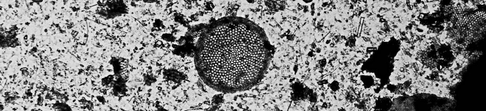
With slide on the microscope stage, take deep breath, focus, and happy
happy I
am. There amid a crowded field of quartz, little boulders, and
undefined debris
is
one large less than perfect centric diatom. Scanning further I found
additional centric diatoms, lots of fragments of centric diatoms,
silicoflagellates, and even a foraminiferan. I was very excited and
pleased.
Many months have now passed and I am still very pleased with my
discovery and I have learned much about separating the wanted diatoms
from the unwanted debris. A sieve is an essential tool in this process.
I had some success with swirling and decanting to separate fossil
diatoms but nothing worked as well as the sieve. An important first
step is to place a small sample of diatomite into a container with
distilled water and subject this to cycles of freezing and thawing,
more cycles seem to be better, roughly 20 to 30, than fewer, 10 cycles
or less.
This cycling does an
excellent job of breaking down the diatomite and freeing the fossil
diatoms before sieving. I have also used hot acid, muriatic acid, to
further separate
and
clean the frustules, silicious shells, after the freeze thaw cycles.
Today I have about 10ml of cleaned material, about
10% of this is diatoms the remaining 90% is fragments of diatoms,
spicules, silicoflagellates, fragments of radiolaria, but no
foraminifera, the
processing apparently
destroys these. Of the 10% that I would describe as usable
material 95% are centric diatoms and a large percentage of these are
Cosinodiscus and the remaining
fraction is a lot of fun to explore.
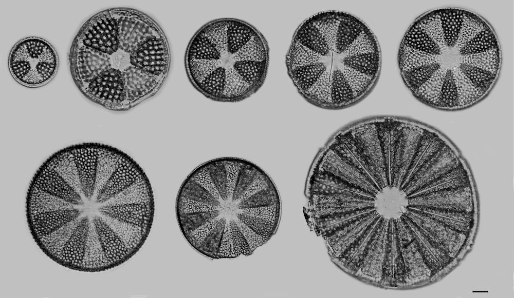
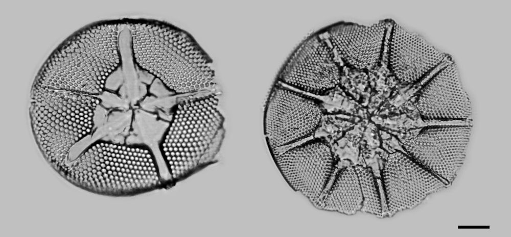
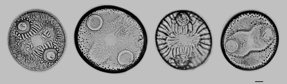
The following images are from the collected diatomaceous earth. The bar
in the lower right corner is the standard, perhaps traditional,
0.010mm.
The images were taken from strewn slides of my manufacture using ZRAX
as a mountant. The diatom images were cut out of their
original frame and pasted onto the neutral background using Paint Shop
Pro.
Genera Actinoptychus
Genera Asteromphalus
Genera
Auliscus
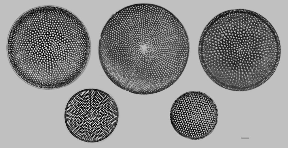
Genera Cosinodiscus
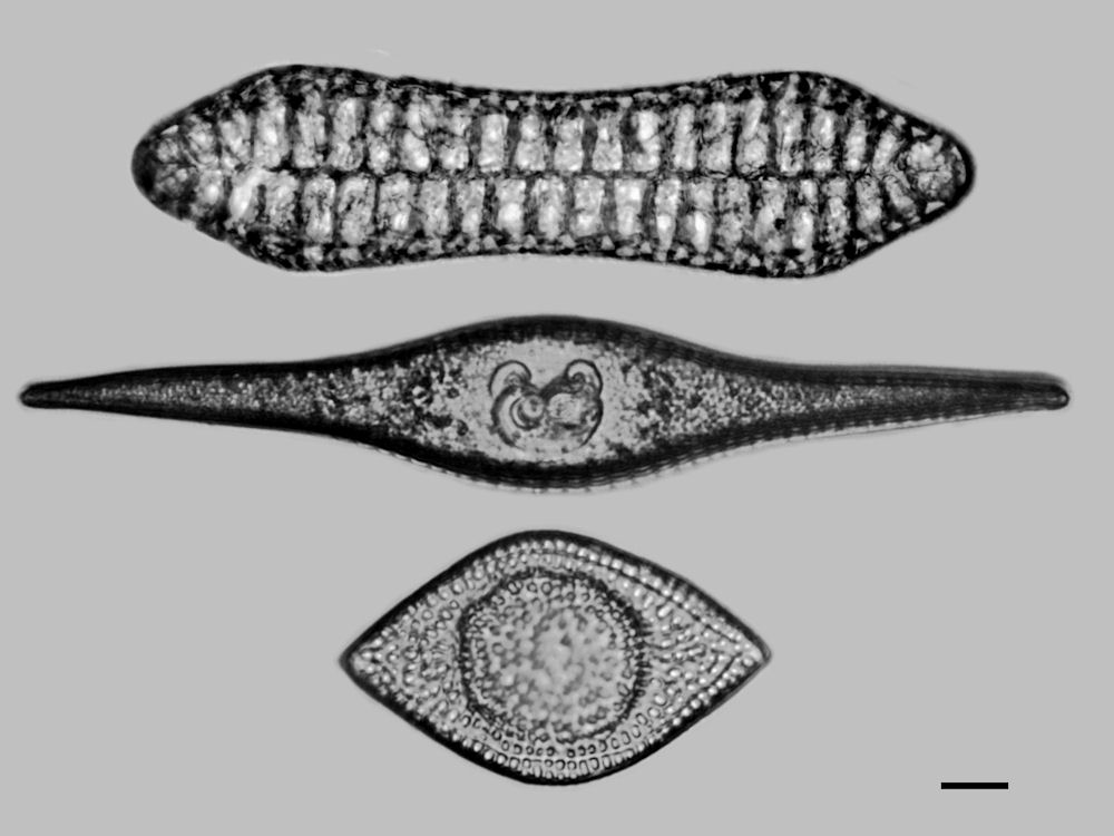
Genera
top
to
bottom
Entopya,
Rutilaria, Craspedodiscus
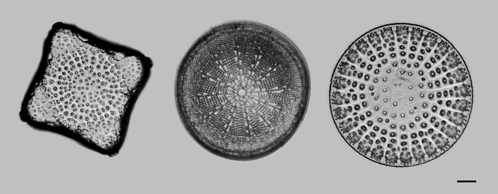
Genera
left
to
right
Triceratium,
Actinocyclus, Stictodiscus
I am very fortunate to have a copy of "Miocene and Pliocene Marine
Diatoms from California" by Walter W. Wornardt, Jr. A 1967 publication
of the California Academy of Sciences. This 108 page paper has become
well thumbed as it is the exact document needed by an amateur working
with Miocene age fossil diatoms of California. My second best source
for information on California marine diatoms is a book in pdf format
currently available from the Scripps Institute of Oceanography "Marine
Plankton Diatoms of the West Coast of North America" by Easter E. Cupp.
A third source that is invaluable to my current endeavor and in
diatoms in general is "The Diatoms Biology & Morphology of the
Genera" by F. E. Round, R. M. Crawford, and D. G. Mann. This book is
currently available and for anyone interested in diatoms this is a must
have addition to their personal library.
Now that I have some experience separating diatoms from the
collected diatomite I wonder how to separate and collect the
Foraminifera from this material? It seems there is always another
interesting challenge just around the corner. As Holmes would say to
Watson "The game is afoot".
Comments to the author are welcomed.
Editor's note: Thanks to Howard McPherson for providing a link to the Cupp reference.
Microscopy UK Front Page
Micscape
Magazine
Article
Library
Published in the April 2010 edition of Micscape Magazine.
Please report any Web problems or offer general comments to the Micscape Editor .
Micscape is the on-line monthly magazine of the Microscopy UK website at Microscopy-UK .
© Onview.net Ltd, Microscopy-UK, and all contributors 1995 onwards. All rights reserved. Main site is at www.microscopy-uk.org.uk .Published in the March 2010 edition of Micscape Magazine.