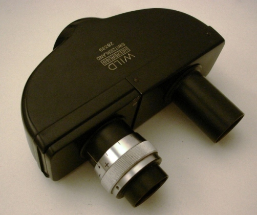
Introduction
This story started a few
weeks ago, when I (re)-entered into the Wild M20 addiction. Browsing
through the Wild M20 resource website, I was appealed by the following
note on the binocular head:
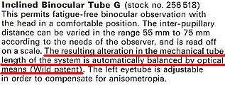
That made me notice that
indeed the interpupillary distance was sent on the M20 by sliding the
left eyetube, while the right eyetube was unaffected. This is quite
unfamiliar, most binocular heads compensate for the interpupillary
distance by varying symmetrically
both eyetubes. How is it possible to maintain focusing by the objective
in the same plane respective to the oculars, while the objective-ocular
distance is varied? How is this specially patented automatic balance performed ?
I immediately wanted to know
more about this (as you're reading this, you already know my geekiness
is clearly above average). My first attempt was to browse the web.
Although the M20 is a famous, well-documented stand, I couldn't find
anything about the binocular tube internal construction. Of course
then, the natural idea is to go by yourself and unmount the item.
However, that goes against one of the major rules in microscopy : "if
it ain't broken, then don't fix it". I don't consider unmounting an
optical piece that doesn't have any trouble is a good idea. Chances are
high, either to bring some dirt or (much worse) to misalign some parts
while remounting.
Finally, I purchased a second
M20 stand (because I wanted its Fluotar objectives and wide-field
eyepieces). The binocular head of this second M20 had some
severe-looking troubles: anti-reflective coatings were attacked, and
the optics of the left eyetube were showing some signs of delamination.
So I came out with an optically imperfect bino, that was enough to
clear-off the aforementioned rule. Hereafter, I will thus discuss about
the internal optics of this defective bino' head, and check about
possible implications towards degrading image quality.
Internal
optics - automatic balance revealed
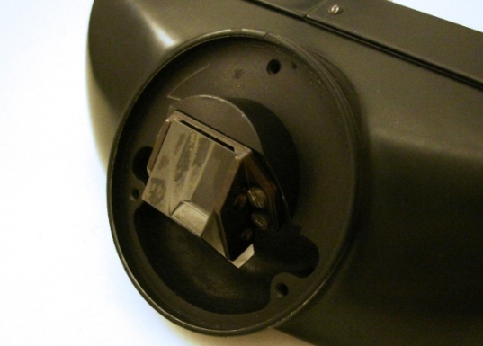
Unscrewing the binocular head
is quite straightforward. The first optical element is a prism to allow
for inclined vision, and is quite usual in microscope tubes.
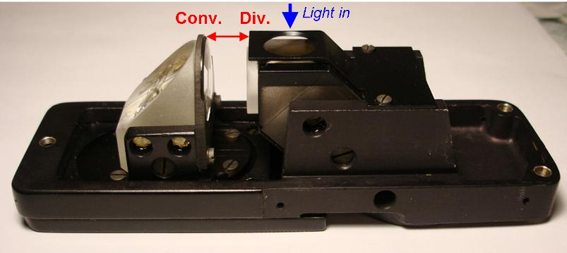
The main optical element is
depicted in the figure below. I hope my annotations will make the
optical path easy to understand for everybody. So here is the secret
into the so-called automatic balance:
a diverging lens fixed to the 50/50 beamsplitter is followed by a
converging lens (almost same curvature). Varying the distance between
these lenses compensates the longer optical length by refocusing the
image formed by the objective. This is a clever construction, yet I
consider the prism-based system (Zeiss) or the symmetrical translation
(Leitz) are intrinsically better. Maybe Wild was coerced into that
design because of patent issues?
How far does delamination in the binocular
head influence image quality ?
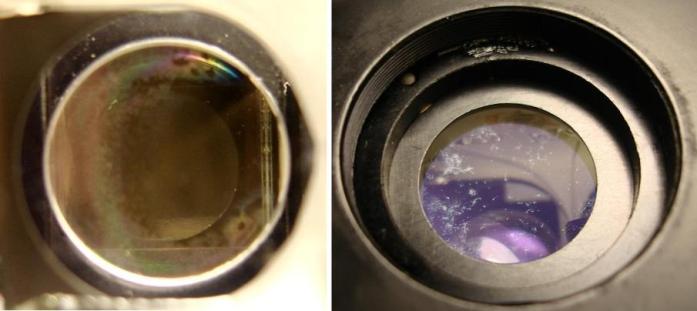
Looking at the internal state
of the optics of the left eyetube, some troubles are apparent: the
diverging lens glue to the prism is strongly delaminating (image below
left), while the anti-reflective coatings also show signs of wear
(image below right, these are not due to bad cleaning procedures, it's
rather the coating that is peeling off).
Can this be fixed ? I'm afraid the answer is no. For the
anti-reflective coatings, one feasible solution would be to remove the
coatings, but it would be hard not to scratch the glass without access
to ultra-strong (hydrofluoric) acids. For the delaminating lens, even if
the lens and the glue can be removed, I don't see how it would be
possible without specialised equipment to (i) avoid any air bubble
while gluing again the lens and (ii) centering all the optics. So
another rule applies here: "if you don't know reasonably how to fix it,
then don't go any further".
The second question now comes to whether all this really affects
imaging, and to what extent? To check this, I used a Wild Fluotar
40x/0.75 phase objective, and my modified Brunel DCM130 USB camera (I
removed all projection optics in the DCM130 to keep only the CMOS chip,
that is directly inserted in a image plane by the objective. Strictly
speaking, the tube length here is close to 180mm, and doesn't meet the
specifications for the objective, but as all this is futile anyway, I
don't really care.) I'm not sure this objective choice is the best, I
wanted to take a high-NA objective that would be more sensitive to
additional aberrations.
The figure below illustrates the different tests undertaken. The sample
is from Klaus Kemp's excellent 8-form diatoms test slide. The images have
been resized, and contrast slightly modified to better illustrate the
discussion.
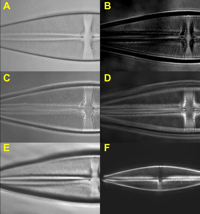
Conditions:
A- bright field, incoherent illumination, aperture iris open to 100% of
objective NA.
B- bright field,
coherent illumination, aperture iris to 5% of objective NA.
C- bright field,
partially coherent illumination, aperture iris open to about 15% of
objective NA.
D- Phase contrast,
annular ring for 40x.
E- Annular oblique
illumination, incidence from the top.
F- Dark field, this
one is taken with Wild Fluotar 20x/0.60.
The effect of
delamination in
the eyetube is only to be seen in case B, and to a minor extend in case
D. It corresponds on the darker zones on the left of the images B,D. In
that cases, resolution is not noticeably affected, it is mostly the
background that turns inhomogeneous.
Conclusions
Delamination in the internal
eyetube is shown to have no noticeable effect on most illumination
conditions. To see any effect, the illumination has to be turned into
strong coherence (small angular divergence of the light illuminating
the sample). In that latter case, it is mostly the background that
turns inhomogeneous. This isn't very detrimental for digital imaging,
as a background can always be subtracted. Fortunately, there is a right
eyetube that does not have these supplementary lenses !
Thanks for reading.
Comments to the author, Jerome Wenger are welcomed !
Microscopy UK FrontPage
Micscape
Magazine
Article
Library
Published in the April 2010 edition of Micscape Magazine.
Please report any Web problems or offer general comments to the Micscape Editor .
Micscape is the on-line monthly magazine of the Microscopy UK website at Microscopy-UK .
© Onview.net Ltd, Microscopy-UK, and all contributors 1995 onwards. All rights reserved. Main site is at www.microscopy-uk.org.uk .Published in the March 2010 edition of Micscape Magazine.