|
New Old Slides: Part 1 by Richard L. Howey, Wyoming, USA |
When in the title, I speak of old slides that are new, I simply mean that they are new to me, not that they are forgeries or fakes of more recent times but, such things do exist and one should be a bit cautious especially when rather extravagant claims are made about some tatty slide that looks like it was made 150 years ago and suffered smoke, rain, and hail damage. They may be being mass produced by some devious, mad graduate student who failed the basic Biology course lab.
Usually at least once a week, I go on eBay to look at the offerings of vintage and antique microscope slides, partly to try to find items of interest and partly so that I can have tantrums about people who indulge in egregiously high bidding (that is, they outbid me on a slide I wanted). This parenthetical remark is not altogether true in that I have virtually no interest in the photo reduction slides of scenes and people which routinely sell for over $100 and, on rare occasions, for several hundred dollars. There are moneyed (or mono-eyed) collectors who are obsessive about acquiring such slides. Chacun à son goût! (which, as everyone knows, means “My horse has gout!”). Also there are small collections of diatom slides that come up for sale fairly frequently and the fraternity of diatomists can be quite frantically aggressive in competing for such collections, even ones that are not particularly distinguished. Over the years, I have acquired some very nice diatom slides either through bidding or as gifts from friends and I much appreciate them, however, in my dotage, I am not inclined to pursue such items with much vigor unless they have some special aesthetic or idiosyncratic character. Recently, I came across a nice late 19th Century slide with 2 specimens of Arachnoidiscus on it. Ordinarily, for a single slide, I am conservative and reluctant to pay more than $35 or $40 for a very good late 19th Century slide unless it is truly extraordinary. This particular slide sold for over $75. It was very nice, but a bit beyond my economic and psychological budget. No problem and I hope the person who bought it will derive real pleasure from it. However, a slide with a variety of sponge spicules from a hexactinellid (glass sponge) came up for sale and, since this is a group of sponges which I have been and will continue to write about, it was of special interest to me; so much so, that I was willing to break my fiscally conservative pound barrier (yes, the dealer was British) and I bid $65 only to discover a day later that someone had out bid me at $104! How dare he or she! He or she deprived you (or saved you from) another one of my essays on spicules. Just this afternoon, an auction for a Watson 1870 arrangement of Synapta spicules, which I had been watching closely, sold for $569! It was indeed a very fine slide and the astonishing thing was that the bidder who won the slide had had zero purchases on eBay before whereas the second highest bidder had made almost 12,000 purchases on eBay and my experience is that such opponents almost always get what they want. I have even seen a single slide sell for over $1,000 which strikes me as beyond the pale–just call me Scrooge.
Just last week, someone outbid me on a Victorian slide of a cross section of a rhinoceros hair. Now, this is not the sort of thing that shows up very often and so one can reasonably expect some competitive bidding. However, I suspect, especially in this age of the Internet, that with the right connections, one could get a whole handful of rhinoceros hair quite inexpensively. Unfortunately there are those nasty ethical questions that arise. Was the rhinoceros poached or killed for its horn to provide an ostensibly pure and natural Viagra for wealthy, gullible Orientals? However, even if the animal had not been poached (which would have required a very large pot), there would doubtless still be all kinds of bureaucratic paperwork to fill out and so something that started out at a reasonable price would suddenly become expensive. The solution: befriend a courageous local zookeeper and if the zoo doesn’t have a rhino, you might have to settle for giraffe or okapi hair or a bird-of-paradise feather. One of the delightful aspects of the English Victorians was their insatiable and often eccentric curiosity that led them to seek out and investigate things that most people would never give a second thought to. To understand this remarkable range, all you have to do is look at listings of the offerings of slide makers from the mid-19th Century through the early 20th Century and then compare those with the slides that are available from biological supply houses today. The audiences differ greatly; today microscopes slides are primarily produced for students and most especially educational institutions; in earlier times, the buyers were predominately moneyed upper middle class members and aristocrats or researchers. Often the distinction between the amateur and the specialist was, in those earlier days, of little significance, since there were amateurs who made highly professional contributions.
Prepared slides can be a valuable teaching tool; however, getting students to seek out their own specimens and learn simple microtechnique can stimulate curiosity in ways that prepared slides and kits never can because ultimately the learning process depends heavily on the excitement of discovery.
So, what does all of this have to do with buying slides on eBay? Well, sometimes a slide can stimulate curiosity by leading you to investigate similar material that you have to find and prepare yourself. Some time ago, I acquired a slide of bat hair and since then I have been looking at hair (and even fibers) from a wide variety of animals (including myself), synthetic materials, and even plants. You’d be astonished at how many fascinating hairs can be found on plants, not to mention beetles. So, I have already sketched out an essay (probably several) on hairs and fibers and that was a major reason that I was disappointed in not being able to acquire the cross section of the rhinoceros hair in the eBay auction.
However, another constructive facet in my effort to educate myself (since I retired, I’m home-schooling myself) is my acquisition over the last several months of 3 collections of antique/vintage slides. I am informed that technically for something to be an antique it must be 100+ years old; so, if it’s only 50 or 99.75 years old, it’s only vintage. We do need some criteria but I’ll tell you that old isn’t necessarily better as my creaky old body and quirky memory readily demonstrate. However, this also most definitely applies to slides. Don’t get me wrong; many of the old slides range from very good to superb but, there are others that were poorly made by incompetent amateurs, slides where the mountant has not stood the test of time and has severely yellowed or cracked obscuring the specimen. Yet others, have either been improperly stored or mishandled resulting in cracked cover glasses, broken slides, or displacement of the specimens. Another, unfortunately rather common flaw, results when a cover glass is improperly sealed resulting either in air bubbles or a rampant growth of mold obscuring or, in extreme, cases, damaging the specimen. So, CAVEAT EMPTOR! (The Emperor has no clothes.)
Over the last few days, I have begun sorting through an old wooden box of 75 slides. These are all botanical sections and so far I have only done a quick check on 6 of them. I can’t find any dates but, I would guess that they were done about 80 or 90 years ago and probably for a botany course at a small college. How did I deduce all of this? “Elementary, my dear Watson. I found a date of 1910 on a couple of the slides and on a strip of paper glued across the inside was written the date “1915". The slide label with the date 1910 had “Dennison University” printed at the bottom. So, where does one go but Google? Well, with that spelling, I found a Dennison Building on the campus of the University of Michigan at Ann Arbor. It is quite modern and devoted largely to mathematics, physics, and astronomy. The other option that appeared was “Denison University”, a small liberal arts institution founded in 1831 and located in Granville, Ohio.
This is no inexpensive local school. For 2011-12 fees, tuition, room and board cost a bit over $50,000 for the academic year.
There are 3 sheets of paper which accompany the box and include a partial listing of the specimens. These are handwritten (hand printed, actually) in pencil, with numerous misspellings and letters so crudely printed as to be indecipherable. At the top of 2 of the pages are written “MAKER: M.E. Stickney” and “CHECKER: D.M. Stickney”. So, perhaps we have an older brother who is a lab assistant reporting on his brother’s work or more likely a father who is a botany teacher who tells his son: “I don’t care if you are allergic to pollen and hate plants; you’re going to learn to make slides of botanical sections.” The only thing wrong with that is that Malcolm Enos Stickney went on to become a distinguished botanist. He got his B.A. and M.A. at Harvard and then did additional graduate work at the University of Wisconsin and at the Marine Biological Laboratories at Woods Hole. From 1909 until his retirement in 1940, he served as Professor and Department Head of the Department of Botany at Denison University. He was also a member of the Ohio Academy of Sciences but, I could find no mention of a D.M. Stickney. In any case M.E. Stickney contributed to the Herbarium at Denison and after his retirement spent a good deal of time working on ferns.
Five of the 6 slides I’ve looked at are quite decently done and so I look forward to examining the remainder. The other 2 boxes pose some different sorts of problems. One contains 72 slides and are identified on the paper index list glued into the inside lid of the box. However, on cursory examination, I have not yet been able to discover any coherent relationship between the labels on the list and the labels on the slides.
The third box of 50 or so slides consists again of botanical sections and I have already told you that though I find them fascinating, this is not one of my areas of even moderate misinformation and this collection poses a special problem. There are no labels on the slides, there is no listing in the box, and there are no handy, informative extra sheets listing the contents. Identification-wise, there is zilch, nada, rien, nichts, so I’ll leave this box until last and maybe by that time, I’ll have learned something about identifying these sections, but don’t count on it.
So, let’s start with the first box that reportedly had 75 slides in it. By the way, my aggressive bidding allowed me to acquire this collection for the sum of $31 plus $10 shipping, so this is a winner on a very nice scale. Furthermore, as I worked my way through the box, I discovered a significant number of instances where 2 slides had been placed in a single slot, so as it turns out there are 115 slides rather than 75.
I noticed slides dated 1908, some 1910 which are thus genuine antiques but, there are some others dated 1913 and 1915, so I guess technically these are vintage. However, since they are so close, I am tempted to coin the them “vintique’ to describe them.
This afternoon I went back to feeding the scientific names written on the slide labels into Google and solved 2 mysteries. (I think I might have made a good Victorian detective.) The first mystery I solved is the name of the institution. I found a slide dated 1914 with “Denison University” printed at the bottom. Oops! Apparently earlier, someone at the university or a hired hand in a printer’s office added an extra “n” and seemingly the university decided “Oh, what the hell, they are just microscope slide labels. Let’s use them up and when they’re gone, we’ll make the correction.” As I mentioned, Denison University is a small and expensive liberal arts institution, but was nonetheless frugal.
Now, to the second mystery. You remember Professor M.E. Stickney. In going through the slides, I discovered that he made many of them and while they are not papered or in any way elaborate, they are very well-prepared and well-stained sections which suggests that these were made as research slides. Nonetheless, his handwriting was awful and, on that basis alone, he could have qualified to be a physician. Some of the slides are of different stages in the development of the same plant and I came across several that were labeled (or so it seemed to me) “Lavix”. Google kept coming up with “Plavix” and I was fairly sure that that would be anachronistic. Eventually I thought, what if that “v” was a sloppily scribbled “r”? The result: up popped “Larch”, a common and well-known tree—a botanical, not a pharmaceutical subject.
There is another wrinkle in dealing with old labels; spelling of genera and species change, there are problems of synonymy and duplication and perhaps, worst of all, names that after a century have simply disappeared from usage. Also, if there are popular and/or local names rather than scientific names then, for a single plant, you might have a dozen, often descriptive, names. Three additional problems crop up: 1) one slide of a given species might have a section of a root, a second one of a stem, a third of a leaf, a fourth of a flower or berry. If these are not specified, then you may have difficulty integrating such sections into your conception of structure of a given plant. 2) Often you’ll encounter cross (transverse) sections but, sometimes you’ll have longitudinal sections and in some instances they will be singular and, at other times, serial thus presenting you with half a dozen or more slices. This can be helpful if you have both transverse and longitudinal sections on the same slide. 3) Then there is the problem of fixatives and stains. Unless particular slides have been discussed in scientific papers, such information is rarely available. Fortuitously, on a few of these slide labels, I have found notations such as “Saf. and Haem.” which is helpful because it tells us that the section(s) were stained with Safranin O and Haematoxylin.
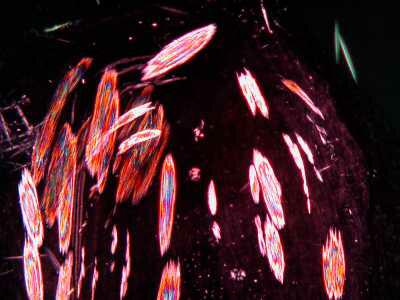
I can hear you mumbling (or if you’re a Type A personality–yelling)–“Come on. Come on. Where are the pictures?” Well, if you’re going to take that attitude, I’ll just make you wait until next month for them (Just kidding.) But, hang in there for another minute or two. I need to tell you something about the way I’m going to present the images, so I don’t have to keep repeating myself, repeating myself, repeating myself. To the delight of microscopists, many parts of plants are birefringent and a considerable number are dazzling under polarized light, especially when you have well-prepared thin sections. In some instances, the staining of the sections enhances the polarization. I will usually present an image of a section using brightfield and then follow it with a polarized image of the same section. In a few instances, I may add a third image where a compensator has been introduced into the polarized light path. Some types of tubules and vesicles, whose function is essentially to transport fluids, show up very distinctly as do certain components which are basically constituted of cellulose compounds. In addition, a large number of plants produce birefringent crystals. For example, you can find them in the leaves of Dieffenbachia and Rhubarb and, in Fuchsia, you can find them in the little “fruits” that develop into seed pods after the flowers have fallen off. Here is an example of Fuchsia from a slide which I made.
Most of the images will be cases of cross sections of plant stems and this presents several problems.
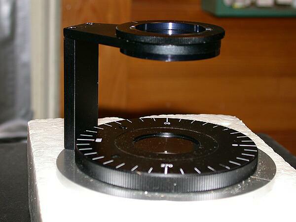
1) Such sections are generally large enough that to get an image of the full section requires the use of a stereo-dissection microscope rather than a compound microscope.
2) This means having a way of achieving polarized illumination for a stereo microscope. I use a handy little device which can be placed directly on the stage. [In a previous article, I have discussed its use in more detail.]
3) In order to align and focus the image for photography, it is frequently necessary to uncross the polars in order to view the specimen and make the necessary adjustments. Then, while holding the stage device firmly in position, one has to rotate the analyzer carefully until the polarizers are again crossed providing a dark background.
4) The focusing, at least with my setup, must be done on the camera screen of the Nikon Coolpix 995 and not through the oculars of my Olympus SZ-Tr Stereo microscope. With brightfield, this is relatively straightforward and the image is most likely to be in sharp focus when there is some scintillation in the specimen on the screen. with polarization, however, it’s not that simple and one may have to take several images to get a satisfactory result.
5) Finally, there is the problem that with very thin sections, as a number in the collection are, adequate contrast, even with staining, may be difficult to achieve with brightfield illumination. Frequently, one can get good results by altering either the intensity of the illumination or by changing the angle of the mirror to get oblique illumination.
So, let’s start taking a look at some of these sections. The first one I’m going to show you is just a brightfield image, since this particular specimen doesn’t exhibit any birefringence but, it gives me an excuse to remember a period from my youth. The plant involved is Sambucus canadiensis or American elderberry.
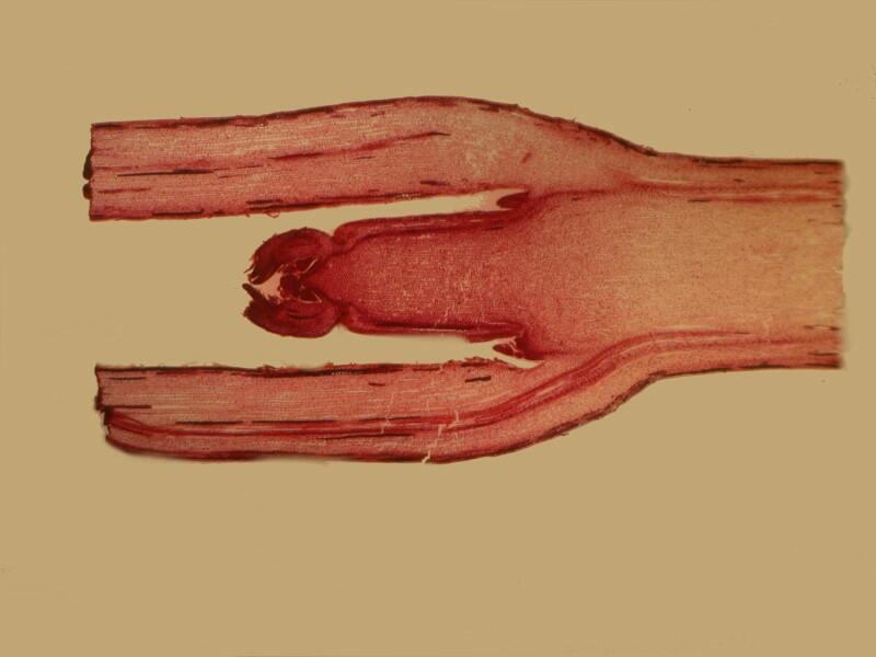
Where I grew up in the Midwest, elderberry grows wild or at least it did 60 years ago. When I was in my mid-teens, on Sundays during the summer, my father, mother, paternal grandmother (who lived with us) and my sister would drive out into the countryside and collect elderberries. This precious cargo was the essential ingredient for the elderberry wine which my parents made and which my grandmother relished while ignoring all other alcoholic beverages except for the occasional nip of blackberry or peach brandy during the winter after the supply of elderberry wine had been exhausted. I felt very grown up because from time to time, as a reward for my help in picking the berries, I was allowed to have a very small glass of this heavy, sweet nectar which I then found magnificent and today would find sickeningly sweet.
Moving on, I’ll bet you didn’t know that the Arum or Calla lily was named after me; its scientific name is Richardia aethiopica (or maybe it was named for Richard III). The images I am going to show you are cross sections of a stem; first, brightfield, then polarized.
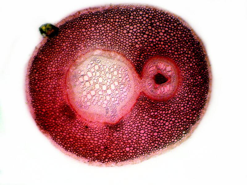
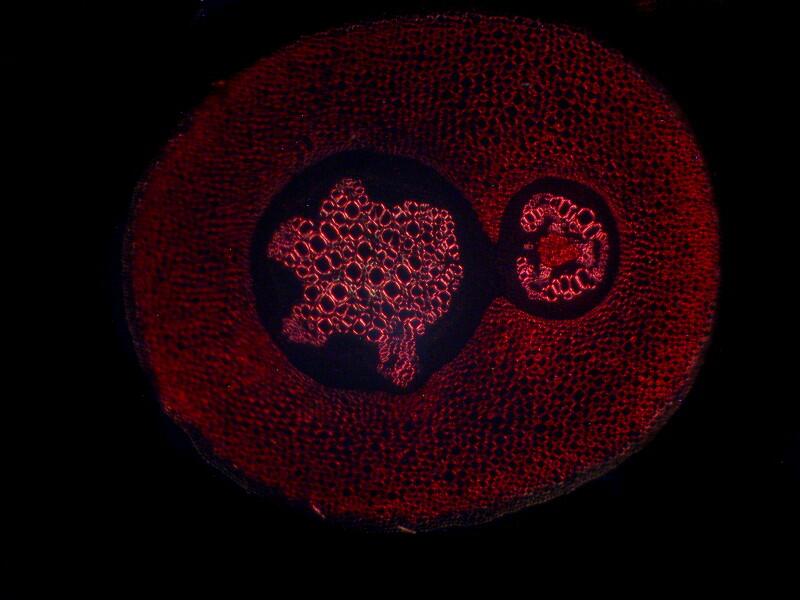
A dramatic difference which underscores the advantages of using as many different techniques to study a given specimen as you have available. In this way, you get a series of perspectives that allow you to gradually develop a multi-dimensional understanding of your specimen and if it has birefringent elements then polarization is often a striking way of seeing elements that you might very well not notice in brightfield even in well-stained sections or organisms.
Let me give you one more example and then I’ll save the rest of Part 2 of this article which I promise will be almost completely images and only very brief comments by me. (I had my fingers crossed when I wrote that last sentence.)
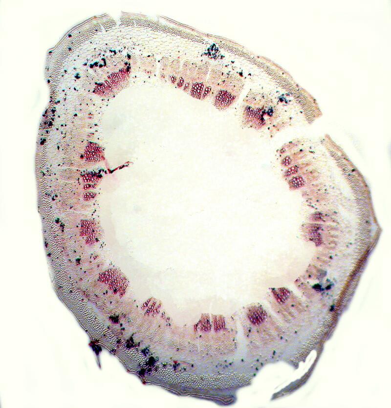
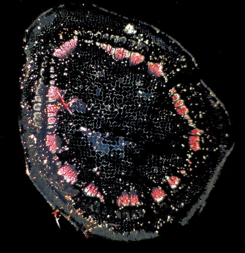
This final example is of cord grass and many grasses and marsh plants provide excellent birefringent material. This one is Spartina stricta and notice how in the brightfield image the center seems virtually empty, whereas in the polarized image, it becomes clear that there is a complex cell structure which can now be observed.
In the next part, we’ll look at some sections of ferns, pine trees, dandelion, corn, butter cup, and Queen Anne’s lace and remember that the images will predominate and I’ll have very little to say. However, I should note then when my wife was proofreading this article and got to that sentence just above, she burst out laughing.
All comments to the author Richard Howey are welcomed.
Editor's note: Visit Richard Howey's new website at http://rhowey.googlepages.com/home where he plans to share aspects of his wide interests.
Microscopy UK Front
Page
Micscape
Magazine
Article
Library
Please report any Web problems or offer general comments to the Micscape Editor .
Micscape is the on-line monthly magazine of the Microscopy UK website at Microscopy-UK .