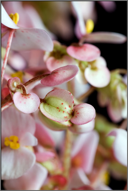
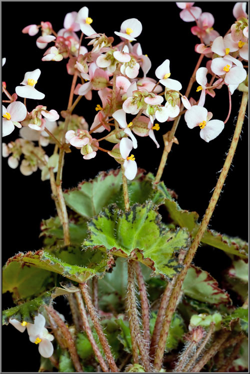
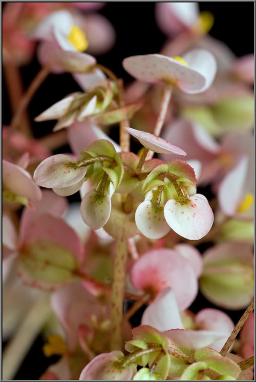
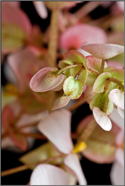
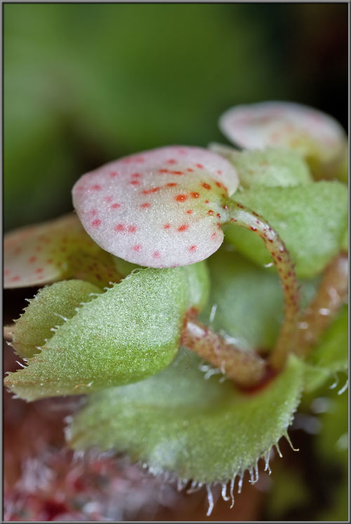
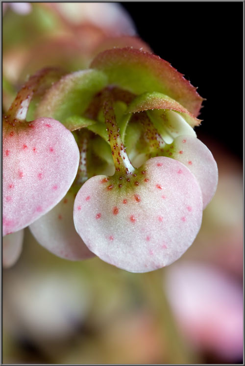
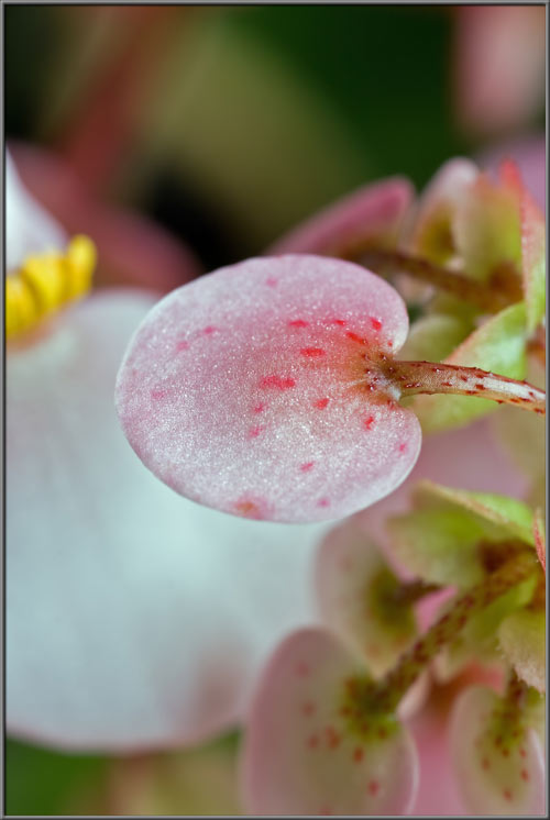
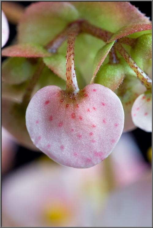
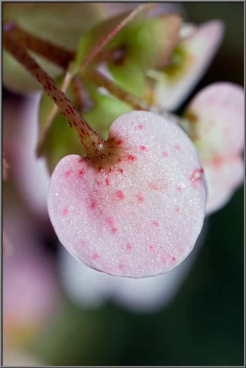
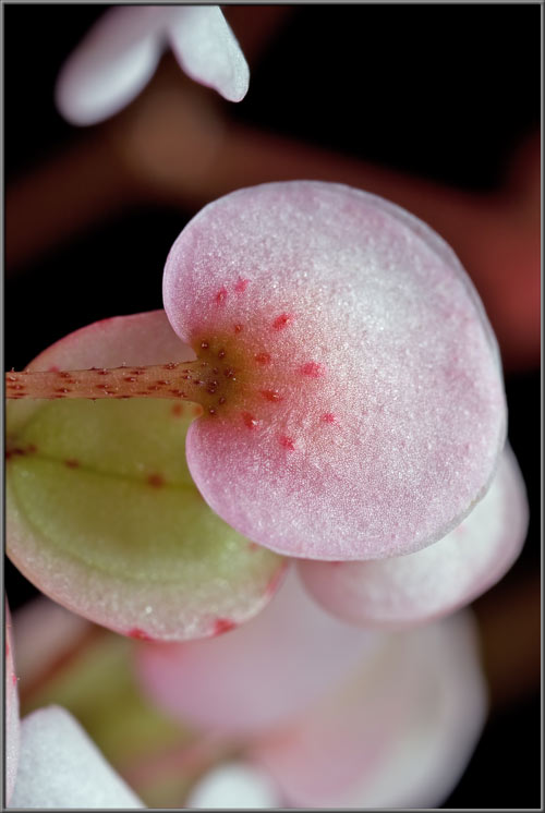
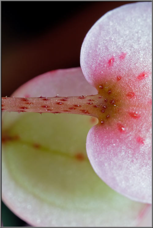
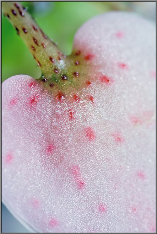
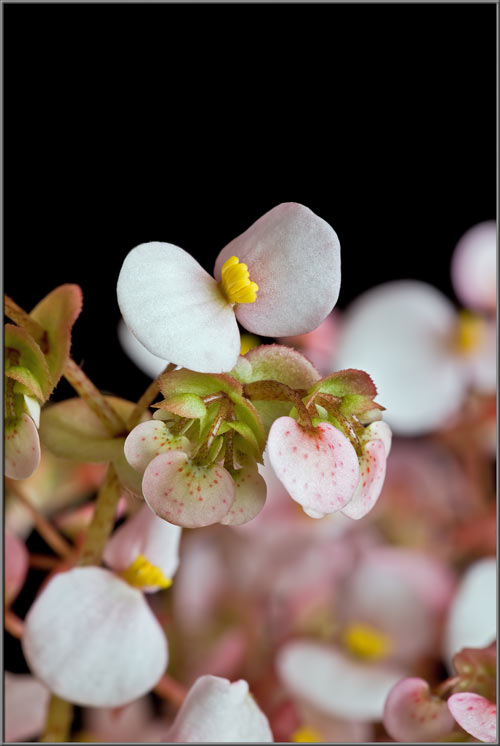
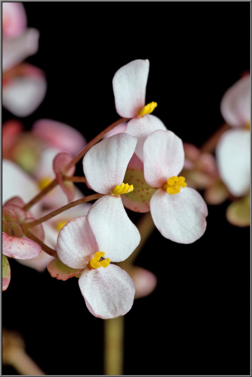
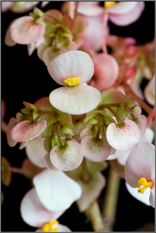
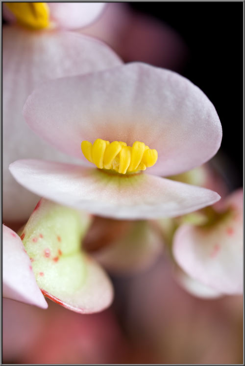
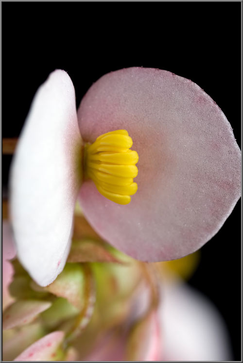
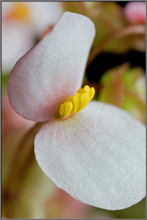
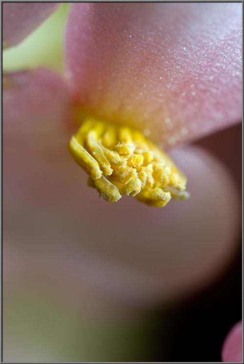
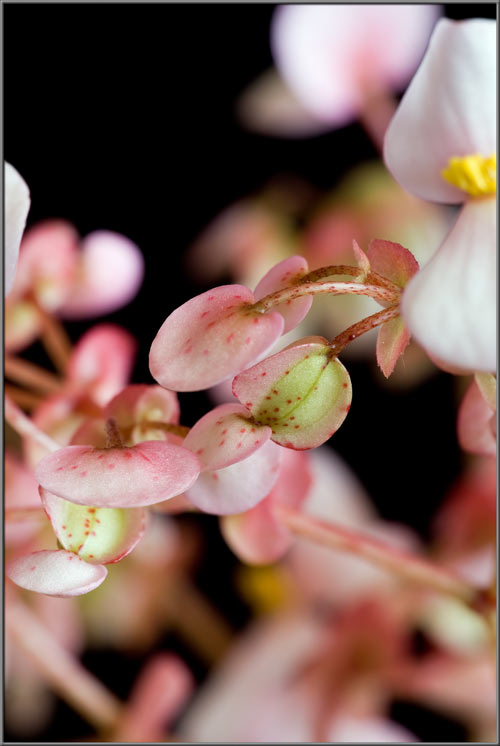
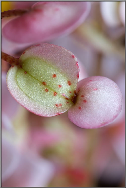
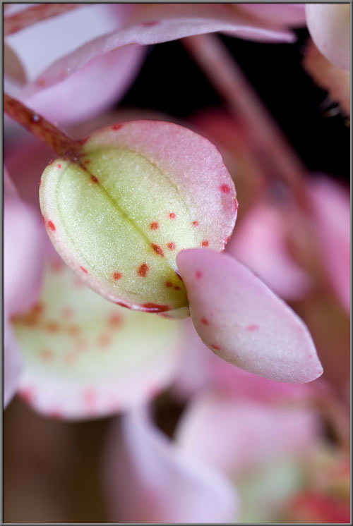
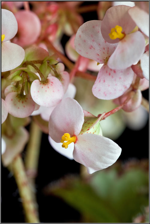
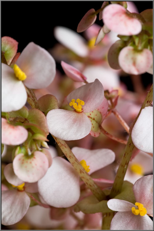
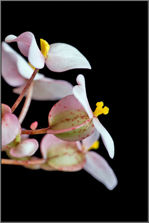
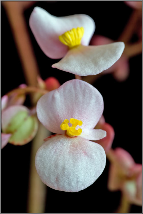
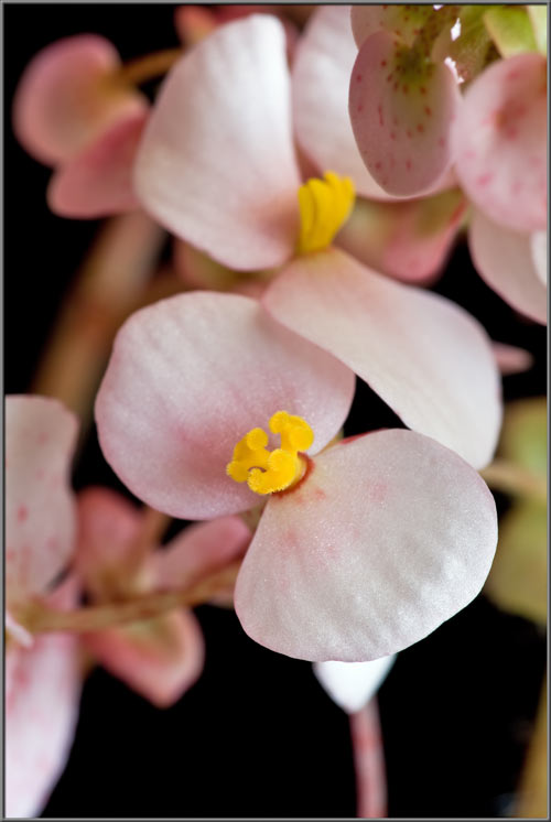
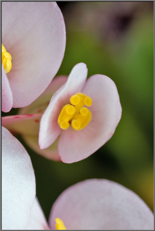
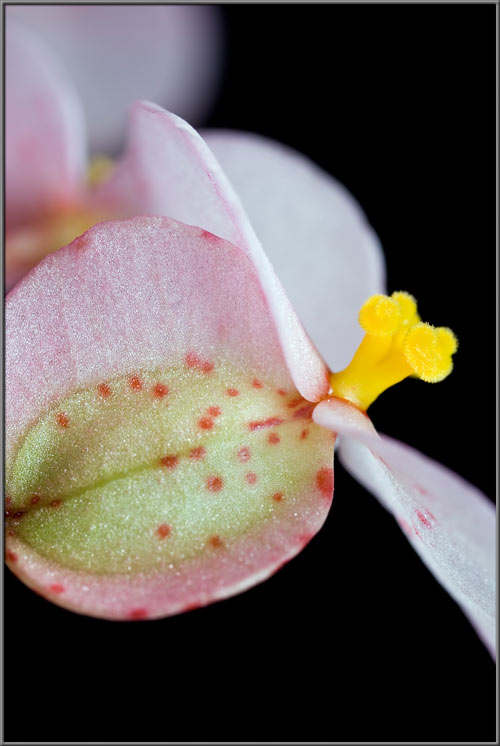
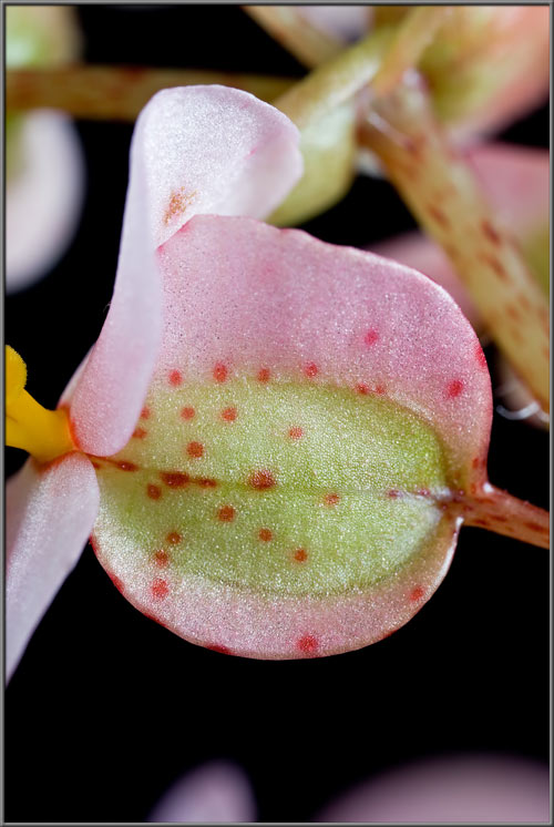
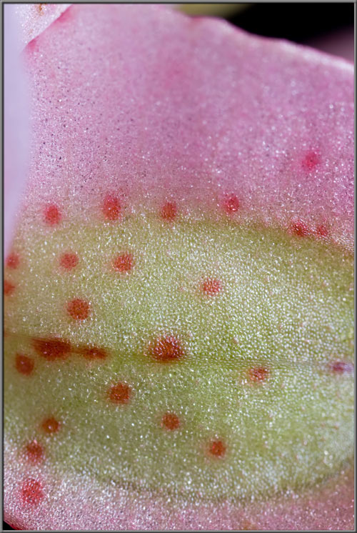
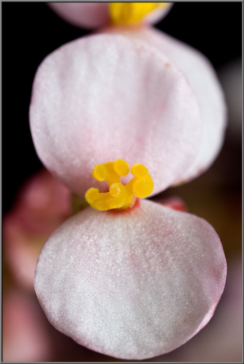
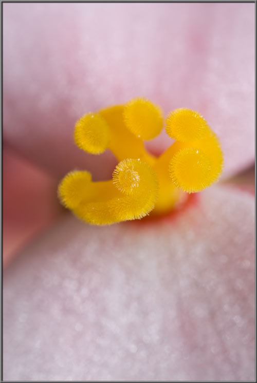
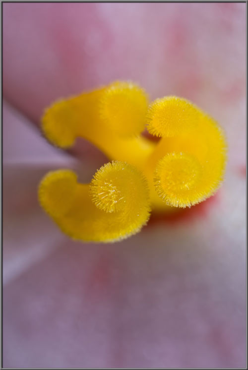
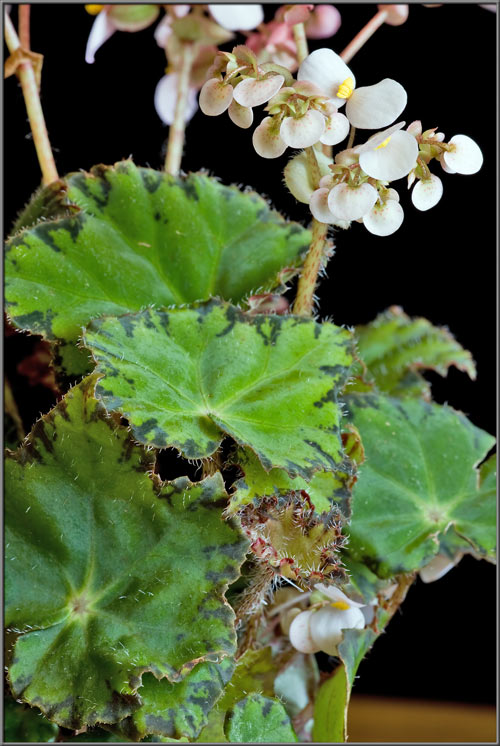
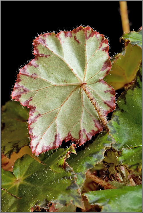
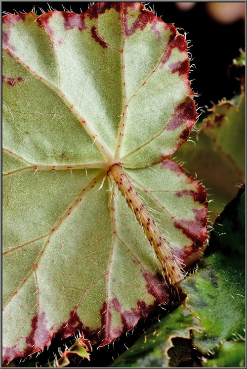
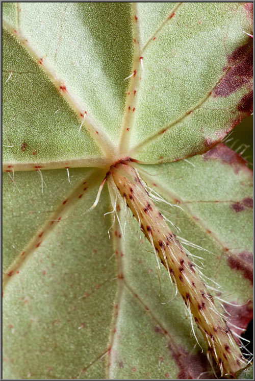
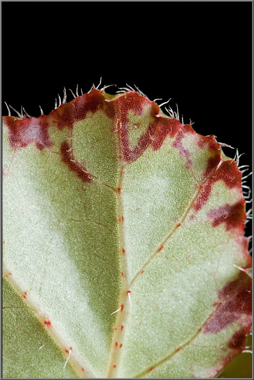
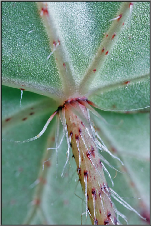
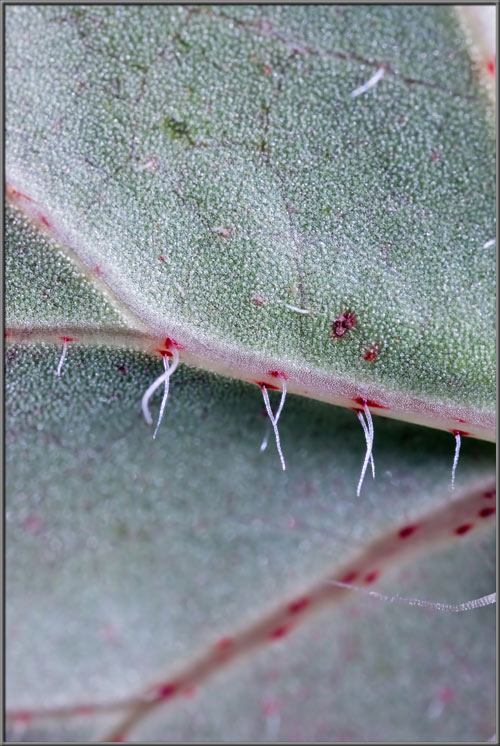
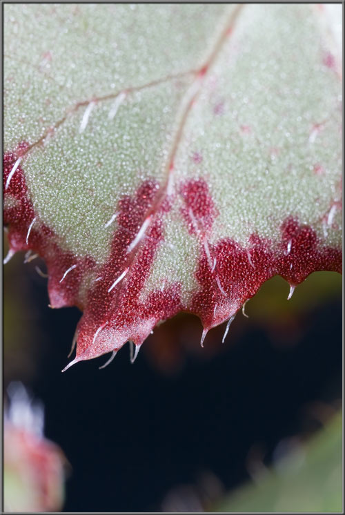
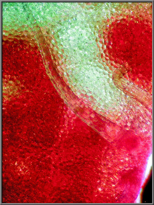
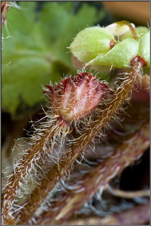
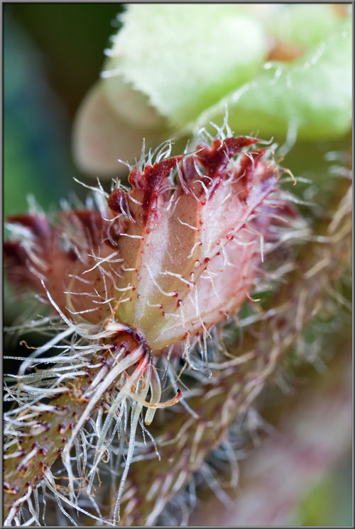
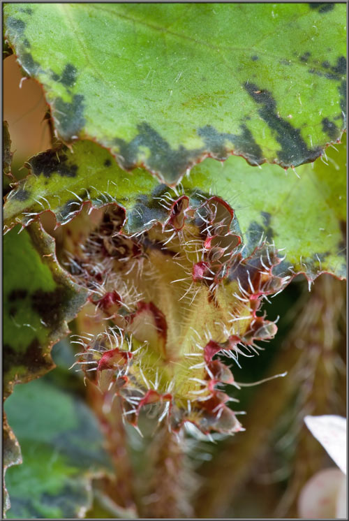
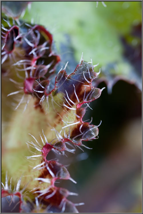
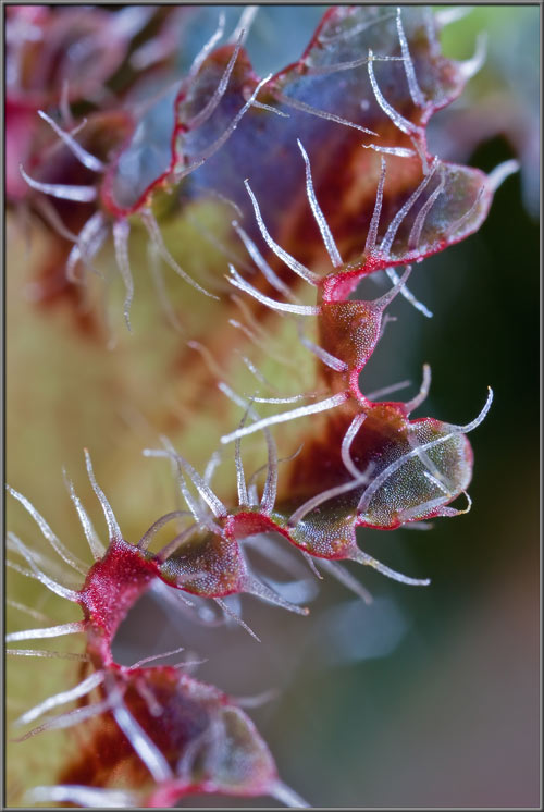
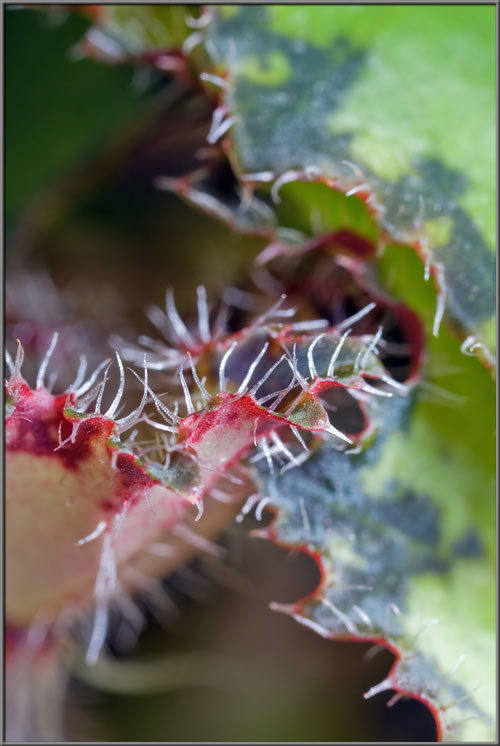
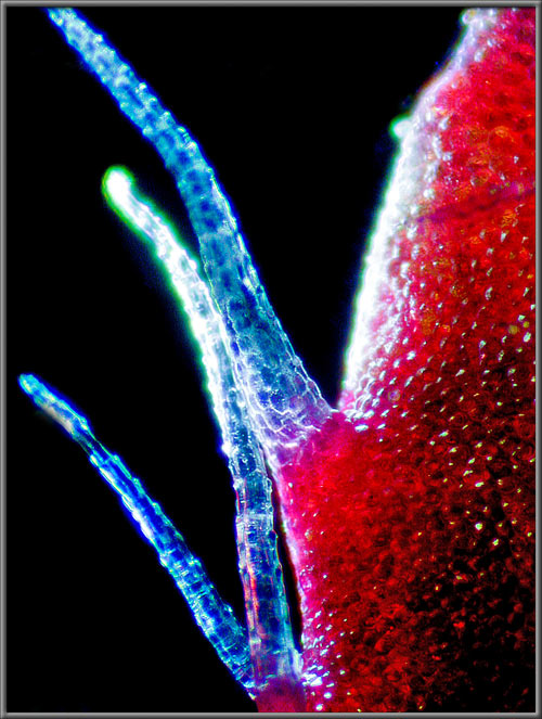
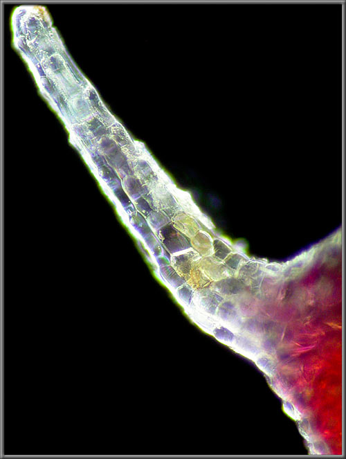
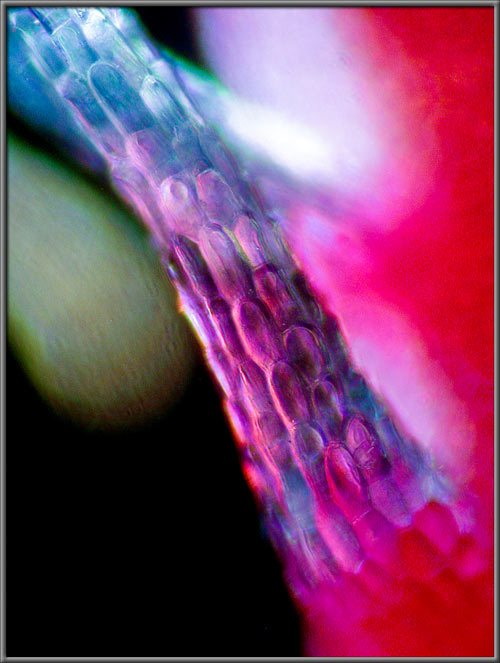
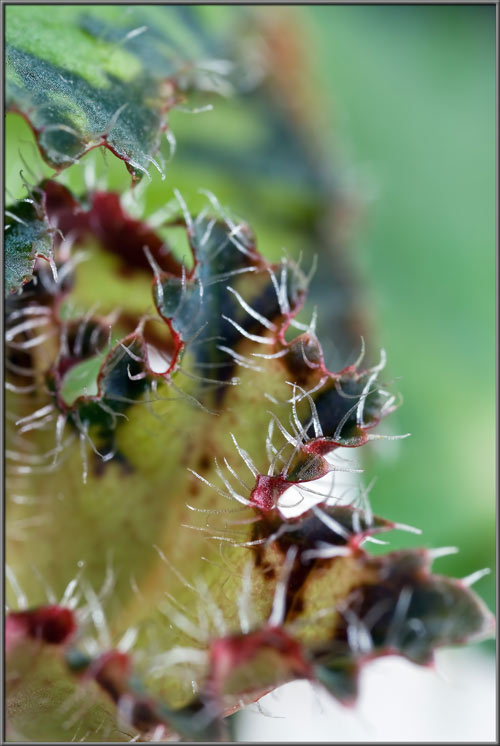
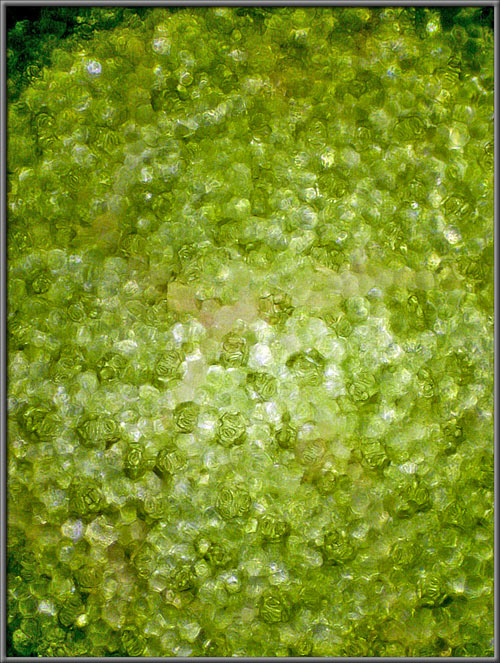
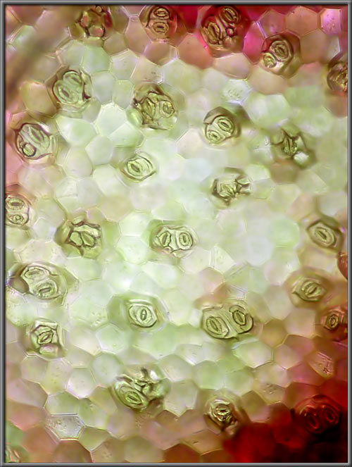
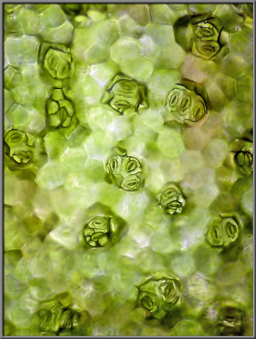
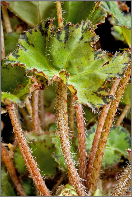
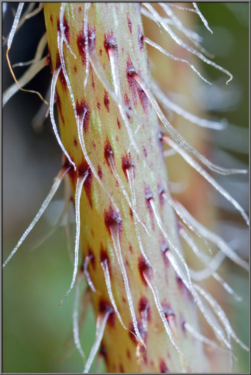
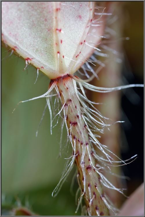
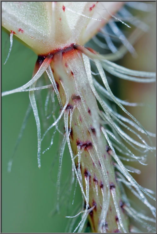
A (Second) Close-up View of a

Flowering Begonia


Begonia x hybrida  

(Family - Begoniaceae)


by Brian Johnston   (Canada)


One year after I completed an article on a flowering begonia, I chanced upon this plant at my local garden centre.  Although it appeared very similar to the first, there were some strange differences that will be noted in this article.  In addition, photomicrographs will provide the closer views that were missing in the earlier examination of the Begonia.  (Note that some of the descriptive text is identical to that of the previous article.)

For the botanical layman, Begonia plants are relatively easy to identify.  Their fleshy jointed stems, showy flowers, and distinctive asymmetric leaves set them apart from other genera.  Unfortunately, an unusual characteristic of Begonias is that species throughout the genus can be hybridized with one another, even if they originated on different continents.  This might seem like a good thing, but it has resulted in a huge number of cultivars to be developed worldwide.  Distinguishing between these hybrids is a formidable task, and so for the purposes of this article, I will simply consider my plant as a ‘typical Begonia’.  Both its common, and genus names were suggested by Charles Plumier, a French patron of botanical research.  Begonia and Begonia honour Michel Begon, a former governor of the French colony of Haiti.

Begonias are monoecious, meaning that they have separate male and female flowers on the same plant.  Male flowers are referred to as staminate, while female ones are called pistillate.  The flower shown at the centre of the first image in the article is pistillate, as revealed by its large winged ovary.  Just to the left of the central flower are two bud-stage staminate flowers which are missing this ovary structure.

The image below shows the plant’s deep green leaves, and multiple flower-heads.  Note the reddish colouration of the plant’s stems.


Let’s look first at the plant’s buds.  It appears as though each flower bud possesses two petals, but in this genus sepals and petals are indistinguishable, and so both are referred to as tepals.  In each bud, the two tepals are closed like the two sections of a clam.  If you examine the images very carefully, you may be able see that some of the clam-like buds are attached to a green base, while others are not.  I wonder what this means?  The answer of course, is that some of the buds are staminate because they are missing the ovary structure at the base of the clam.  In the left hand image all of the buds appear to be staminate.  Several of  the buds seen in the image on the right are pistillate, as evidenced by their light green ovaries.  In most bud groups, there are both staminate and pistillate types.


If we look more closely at the staminate buds, it appears that their surface colouration runs from almost white to a medium pink.  All have a random sprinkling of darker pink dots.  These pink dots are also found on the buds’ stalks.


The plant studied in the earlier article had many bulbous glandular hairs covering the bases of buds, and their supporting stalks.  These glandular hairs are completely absent in this hybrid.


Under the microscope, the petal's ‘spots’ can be seen to be composed of tinted, elongated cells.  The images are ‘false colour’ because the ‘Levels’ function in Photoshop was used to increase contrast.


As the staminate buds open, their two tepals separate to reveal a curved, flattened group of yellow anthers.


Closer views of a flower’s stamens reveal that they are supported by broad, but very short yellow filaments.  Notice the variation in shape and size of the anthers themselves.


The presence of the swelled, winged ovary at the base of a pistillate flower makes it easy to identify.


The side and top views of such a flower show its basic structure.  The unopened bud's tepals, and ovary wings have a light pink colouration, while the ovary itself is a light green.  The ovary’s three wings are positioned 120 degrees apart.


When the flower’s two tepals open in the blooming process, its bright yellow pistil is revealed.


Since the pistillate flower’s ovary is divided into three chambers by its wings, there are three pistils, each with a bi-lobed stigma, and supporting style.


Closer views show each stigma’s two lobes more clearly, and that the three styles are fused near their connection to the ovary.  Notice the bright red line that marks this meeting point.


Notice the prominent spots on the body and wings of an ovary.


Under the microscope, the red colouration appears to be located ‘beneath the surface’.  I suspect that the tinted cells may be located in a layer positioned beneath the surface one.


As we move closer and closer to the elegantly shaped bi-lobed stigmas of a flower, their coatings of fine, hair-like projections become visible.  These fine hairs aid in the capture and retention of pollen grains transported by wind or insects.


The following high magnification photomicrograph shows these hairs clearly.


Begonias are noted for their asymmetric leaves.  Their upper surfaces have approximately 8 rounded lobes.  Note that around a leaf’s perimeter it has a dark, irregular band which is sometimes tinged with red.  (Begonia leaves contain the chemical calcium oxalate within their cells, which makes them poisonous to animals.)


The underside of a leaf is more colourful than its upper surface.  Lighter in colour, with a radial array of prominent veins, the leaf possesses a deep red, irregular band around its perimeter. 


In the earlier Begonia studied, the point of connection of stalk to leaf had a bright red ‘beard’ of long hairs.  In this cultivar however, this beard is almost non-existent.  Both stalk, and leaf edge are liberally covered with fine colourless hairs, with those on the stalk being longer.


Even the radial veins have colourless hairs growing from them. 


The image on the right below shows a photomicrograph of the base of one of these hairs.  It appears to be pink in colour, but it is not.  The hair is transparent and the colour is due to the red spot beneath it.


A mature leaf develops from the hairy structure seen below.  At first the tiny leaflet is more pink than green.


Views follow, taken from above, showing the interior of the partly furled leaf.  The outer edge is bright red, convoluted, and has many hairs growing from it.


If one of the hairs is examined under the microscope, details of its cellular structure can be seen (right hand image).


Higher magnifications reveal the elongated cells near its base.


The green material forming the leaf’s surface is examined next.


The following series of photomicrographs taken with increasing magnification reveals the stomata and guard cells that control gas entry into, and egress from, the interior of the leaf.


The Begonia studied here grows from a unique root structure that looks like a large tuber called a caudex.  The plant’s stems grow from this caudex, which is half beneath the soil, and half above. Multiple stems originate from each caudex.


Four views of stems follow that show their pale yellowish-green colouration, and tiny bright red spots.  The length of the stem’s irregular hairs is very variable but they tend to be the longest near the attachment point of stem to leaf.


A number of strange egg-shaped objects were visible on structures that I examined under the microscope.  Several examples can be seen below.  They look like developing seeds that might be found in the ovary, but this is unlikely, since I did not cut open any of those structures.


The approximately 1500 species of wild Begonias grow in a wide variety of ecological niches, most in tropical and sub-tropical regions.  Only one tropical region is without native species, and that is Australia!  Horticulturalists the world over have developed many cultivars, some to highlight their unusual leaves, and others to showcase particularly colourful flowers.


Photographic Equipment


The low magnification, (to 1:1), macro-photographs were taken using a 13 megapixel Canon 5D full frame DSLR, using a Canon EF 180 mm 1:3.5 L Macro lens.

A 10 megapixel Canon 40D DSLR, equipped with a specialized high magnification (1x to 5x) Canon macro lens, the MP-E 65 mm 1:2.8, was used to take the remainder of the images.

The photomicrographs were taken using a Leitz SM-Pol microscope (using a dark ground condenser), and the Coolpix 4500.


A Flower Garden of Macroscopic Delights

A complete graphical index of all of my flower articles can be found here.


The Colourful World of Chemical Crystals

A complete graphical index of all of my crystal articles can be found here.


 All comments to the author Brian Johnston are welcomed.

Microscopy UK Front Page
Micscape Magazine
Article Library


© Microscopy UK or their contributors.

Published in the April 2013 edition of Micscape.
Please report any Web problems or offer general comments to the Micscape Editor.
Micscape is the on-line monthly magazine of the Microscopy UK web
site at Microscopy-UK  


© Onview.net Ltd, Microscopy-UK, and all contributors 1995 onwards. All rights reserved. Main site is at www.microscopy-uk.org.uk with full mirror at www.microscopy-uk.net .