|
Display by Richard L. Howey, Wyoming, USA |
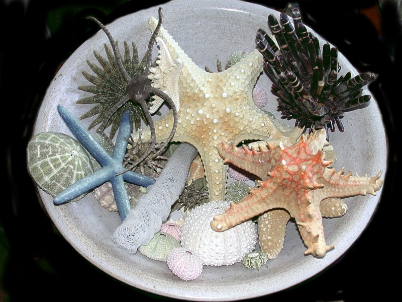
Many people interested in Natural History gradually build a collection of specimens over the years and often some of them are of sufficient interest that we come to feel that displaying a small collection of them might be desirable. I am going to give you an example of one such display. This was done in a large pottery bowl. First, I’ll show you the whole display and then we’ll take a more detailed look at some of the individual specimens.
One of the more striking specimens is the starfish with the red spines. This is Protoreaster linckii.
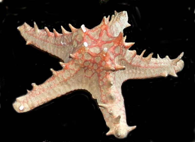
I have another specimen of it in which the color is somewhat better preserved.
However, the real vividness and brilliance of the red can only be seen in a living specimen.
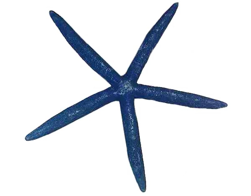
The vast majority of marine animals lose all or most of their pigmentation when they are preserved and some look very drab indeed. There are, however, a few exceptions such as the blue starfish, Linckia laevigata; nonetheless, the coloring in a living specimen is richer and even more striking.
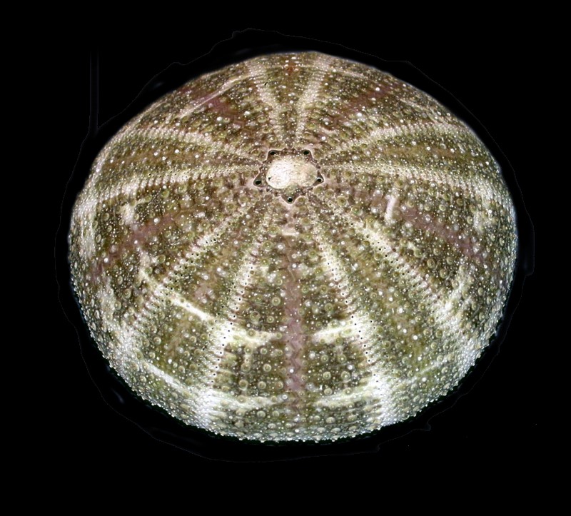
Sea urchins either with spines or denuded tests are always pleasing additions to such a display. A very popular echinoid test is that of the Alfonso urchin, Toxopneustes pileolus.
There is considerable variation both in coloration and in pattern in this genus as you can see here:
However, even more surprising is the fact that from looking at the test there is little in the way of clues as to what the living organism looks like. It is also known as the “flower urchin” and is quite toxic. What look like tiny flowers are actually pedicellariae which are circular when “open” and when disturbed they close and take on a triangular shape. Each one has 3 “jaws” with a sharp hooked barb which can inject poison.
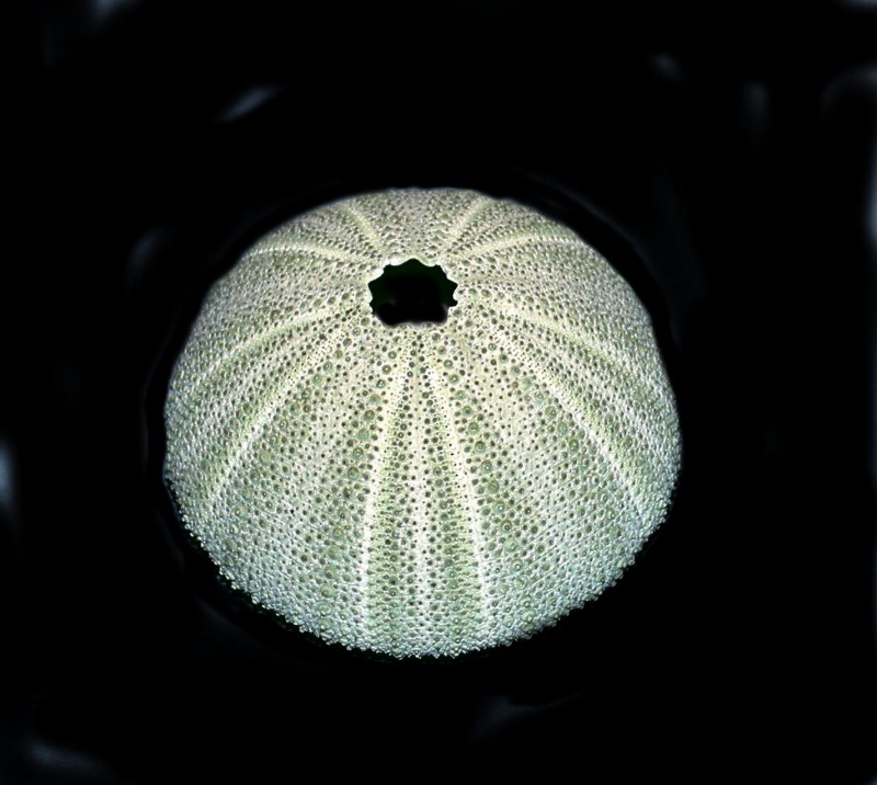
Here is a sea urchin from the bowl of quite a different sort. This is a green urchin, Salmacis spheroides and the minute markings on the test are particularly remarkable when you realize that each little knob is a place where a spine was attached and each tiny hole is where a tube foot extended through.
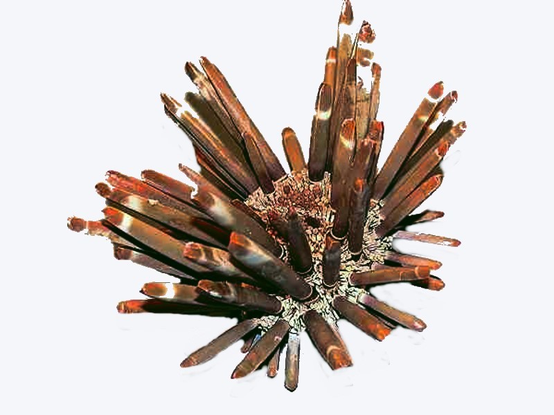
The large club spine urchin from the bowl is a specimen of Heterocentrotus mammilatus. These are tropical organisms and are widely collected and craft shops often sell packets of the spines.
There is a small pink urchin at the edge of the bowl which is an example of another type that craft people and natural history collectors frequently obtain; it is a specimen of Echinometra mathaei.
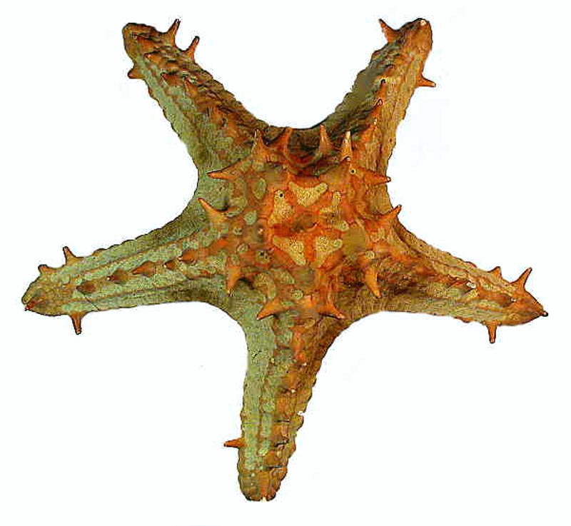
The large tan-colored starfish in about the center is called a “jungle” starfish. There are lots of images of it on the Internet, but I was unable to find any scientific identification of it.
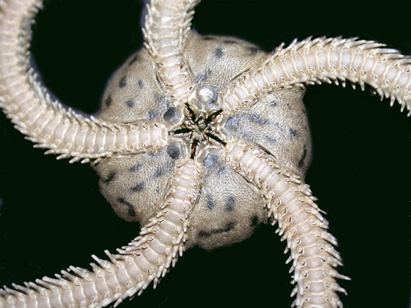
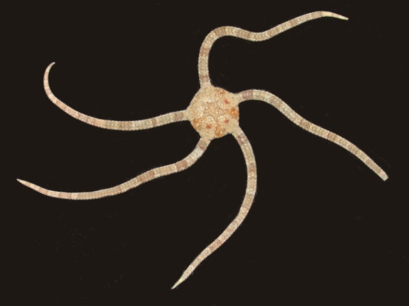
Just to the left of it is a ophioroid or “brittlestar” and a fairly large one as these creatures go. Here is a closeup of the ventral side and you can see the pentagonal pattern repeated in the oral area.
Here is another species (both this one and the one above are tropical forms from the Philippines). This one has retained more of its color. Some species are brilliantly colored. Brittle stars can occur in enormous numbers and certain of them in the ocean bed are sometimes described as “sea gardens” with thousands and thousands of ophioroids snaking across the substrate.
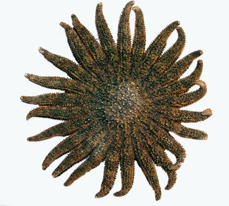
Just under the brittle star in the bowl is a Heliaster or “sun star”. This specimen has 25 arms.
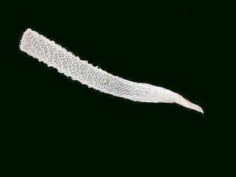
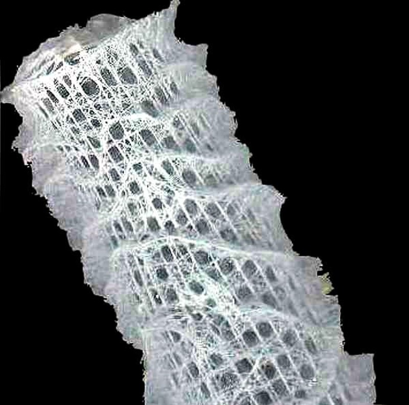
Finally, the long white tube extending our from under the “jungle” starfish downward to the edge of the bowl is the skeleton of the glass sponge Euplectella aspergillium, one of the hexactinellid sponges which have 6-pronged spicules. I’ll show you 2 images; first, the complete skeleton and then a closeup which shows some of the detail of this remarkably beautiful structure.
I have written fairly extensively on Euplectella and you can find 4 of my articles on Micscape on this sponge.
This particular display is not as orderly as it should be since some of the specimens are not very clearly visible. This is, in significant part, a consequence of the help I get from our 3 cats who from time to time like to rearrange some of the specimens.
All comments to the author Richard Howey are welcomed.
Editor's note: Visit Richard Howey's new website at http://rhowey.googlepages.com/home where he plans to share aspects of his wide interests.
Microscopy UK Front
Page
Micscape
Magazine
Article
Library
Published in the April 2017 edition of Micscape Magazine.
Please report any Web problems or offer general comments to the Micscape Editor .
Micscape is the on-line monthly magazine of the Microscopy UK website at Microscopy-UK .
©
Onview.net Ltd, Microscopy-UK, and all contributors 1995
onwards. All rights reserved.
Main site is at
www.microscopy-uk.org.uk .