Fun with graphite and sticky tape. -
The 'what if' 'Friday Night Experiment' that led to a Nobel Prize.
by David Walker, UK
 I've recently been reading about graphene and the simple experiments with graphite and sticky tape which led to the 2010 Nobel Prize in Physics for Andre Geim and Konstantin Novoselov both at the University of Manchester,
UK. The Prize was awarded "for groundbreaking experiments regarding the two-dimensional material graphene". The researchers were seeking reliable ways of making usable and well defined fragments of carbon monolayers to study its properties. The prize winners sourced their first pieces of graphite for further thinning from the discarded sticky tape routinely thrown in a bin which scanning tunnel microscopists used to clean their graphite surfaces. (See e.g. Graphene by Les Johnson and Joseph E Mealey, 2018, Prometheus Books, p.80.)
I've recently been reading about graphene and the simple experiments with graphite and sticky tape which led to the 2010 Nobel Prize in Physics for Andre Geim and Konstantin Novoselov both at the University of Manchester,
UK. The Prize was awarded "for groundbreaking experiments regarding the two-dimensional material graphene". The researchers were seeking reliable ways of making usable and well defined fragments of carbon monolayers to study its properties. The prize winners sourced their first pieces of graphite for further thinning from the discarded sticky tape routinely thrown in a bin which scanning tunnel microscopists used to clean their graphite surfaces. (See e.g. Graphene by Les Johnson and Joseph E Mealey, 2018, Prometheus Books, p.80.)
There is a wealth of information on graphene online (the Nobel Prize site provides a number of background documents). I was interested in mimicking the typical first experiments to observe the results under an optical microscope.
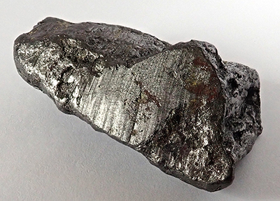
Right: Online procedures suggest that the source of graphite can be from marking a small ca. 1 cm square on paper with a soft pencil. Preferring to use the original source I bought a small piece of graphite for a few pounds from a geology shop on eBay UK. An area has been scraped flat for better contact with tape.
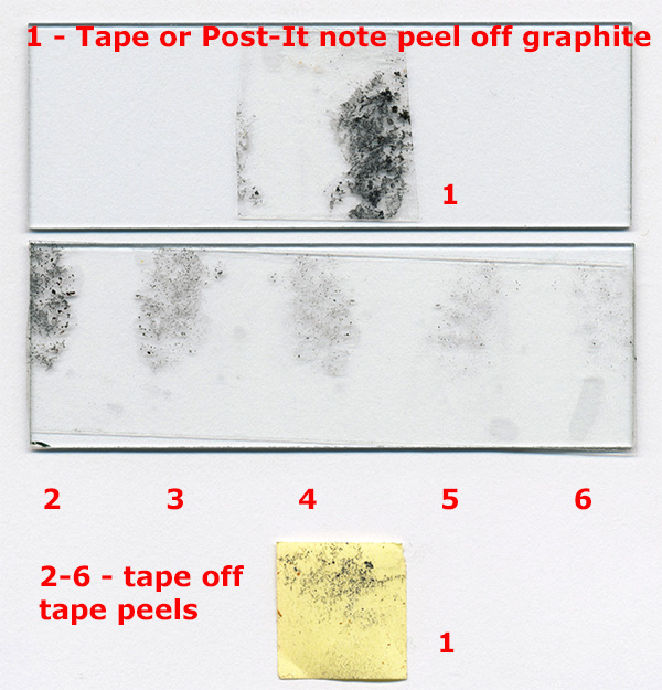
The original procedure describes lifting some graphite from the source using one end of a longish piece of sticky tape (Scotch tape aka Sellotape). Further thinner flakes are attained by pressing the first lift-off onto an adjacent clean piece of the same length tape and repeating. A typical result after six lifts is shown below. Even to the eye it is quite clear that progressively thinner
flakes are achieved to the point of more being transparent. This approach seems potentially more prone to creating artefacts under the optical microscope so tried a Post-It note instead. This does lift graphite off the source as shown below.
The offline and online resources note that graphite becomes transparent for many layers of carbon sheets and well before graphene has been formed. This is what seems to be happening with the example below as even a first lift shows some transparent areas as part of larger opaque fragments. But it is still a simple and neat experiment that can be carried out with any student compound microscope to demonstrate the first steps to a new form of carbon with unique properties and potentially exciting uses which are currently being explored. To determine if genuine monolayers of graphite flakes were achieved, Geim and Novoselov used interference optical microscopy. The flakes when adhered to a 300 nm silicon oxide layer on a silicon wafer showed a change in interference colour as they progressively thinned to a monolayer of graphite (see Johnson and Mealey, p. 82). Could this technique be mimicked by the enthusiast e.g. with epi DIC? Comments welcomed.
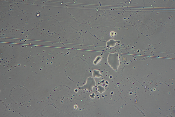
Above. Sellotape gives a potentially confusing image of tape in good contact with the slide and raised areas encapsulating tiny specks of graphite. The above is a control of a clean Post-It note pressed firmly with back of fingernail to slide, pulled off then coverslip added. The control
illustrates the typical artifacts such as these amoeboid-like areas of glue or other deposits but are at a low density. Zeiss 16X Neofluar objective with phase.
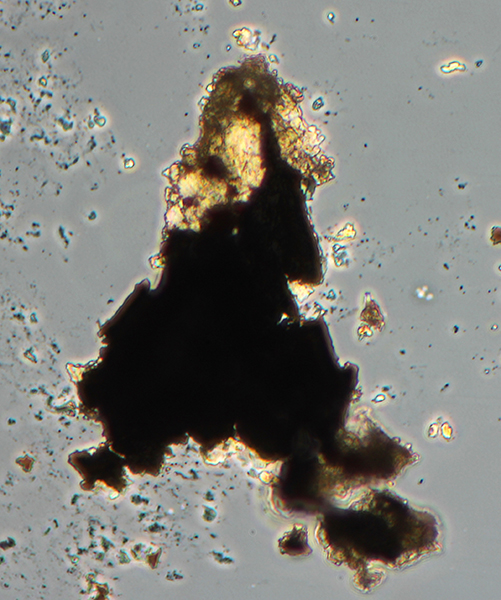
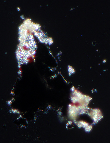
Above. A single lift off graphite using a Post-It note. Zeiss 16X Neofluar objective with phase. There are potentially many isolated transparent pieces of graphite but it is uncertain if they are other debris. The most reliable evidence of thinned graphite to the point of transparency is likely when, as shown here, it is an extension of a well defined opaque fragment of graphite.
Right. The same fragment under crossed polars. The transparent areas seem to show some birefringence as these areas would also be opaque under crossed polars if not.
Comments to the author are welcomed.
©
Microscopy UK or their contributors.
Published
in the April 2020 edition of Micscape.
Please
report any Web problems or offer general comments to
the
Micscape
Editor
.
Micscape
is the on-line monthly magazine of the Microscopy UK web
site at
Microscopy-UK
©
Onview.net Ltd, Microscopy-UK, and all contributors 1995
onwards. All rights reserved.
Main site is at
www.microscopy-uk.org.uk.