| Oblique
and Axial Illumination by
John S. Wojtowicz, US
|
The compound microscope most commonly used by amateurs is
designed for transmitted illumination. Much of the
"unprepared" world is opaque and unless the material
can be cleared and sectioned it is invisible to such an
instrument at higher powers. Magnifications involving the 4X to
10X objectives are usually not a problem as the working distance
of those objectives is great enough to allow room for an incident
illuminator and the optical aberrations introduced by the lack of
a cover slip are negligible.
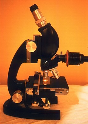 It is otherwise
with high magnifications, and several lighting techniques are
available. The microscope pictured is equipped for axial
illumination. The stand is similar to a biological microscope
with a number of significant differences. The three objectives
8X, 20X, and 40X are corrected for use without a cover glass and
for an optical tube length of 215mm rather than the standard
160mm. Like so much else in microscopy these standards vary with
the manufacturer. There is no substage condenser, just a mirror
for the occasional transparent subject viewed at low
magnification. The illuminator is located between the objective
and the binocular head. The light is reflected onto the subject
by a half silvered mirror located just above the back lens of the
objective. This acts as a beam splitter, allowing some of the
light to be reflected downward towards the subject, yet also
allows the image forming light rays to pass through the mirror.
Thus, the lens itself acts as the condenser. Since the
illuminating and image forming light rays cross, the glare can be
considerable. The mirror can be withdrawn if desired when oblique
or transmitted illumination from a separate light source is used.
It is otherwise
with high magnifications, and several lighting techniques are
available. The microscope pictured is equipped for axial
illumination. The stand is similar to a biological microscope
with a number of significant differences. The three objectives
8X, 20X, and 40X are corrected for use without a cover glass and
for an optical tube length of 215mm rather than the standard
160mm. Like so much else in microscopy these standards vary with
the manufacturer. There is no substage condenser, just a mirror
for the occasional transparent subject viewed at low
magnification. The illuminator is located between the objective
and the binocular head. The light is reflected onto the subject
by a half silvered mirror located just above the back lens of the
objective. This acts as a beam splitter, allowing some of the
light to be reflected downward towards the subject, yet also
allows the image forming light rays to pass through the mirror.
Thus, the lens itself acts as the condenser. Since the
illuminating and image forming light rays cross, the glare can be
considerable. The mirror can be withdrawn if desired when oblique
or transmitted illumination from a separate light source is used.
The illuminator has both field and aperture diaphragms, but
their positions are reversed compared with a biological
microscope. The aperture diaphragm located near the light source
can be used to increase depth of field at the expense of
resolution as in transmitted light instruments. The field
diaphragm is critical for the control of glare. In fact I find I
frequently close it down so that it blocks out much of the
available field of view to get better lighting on details of
interest. This is a particular advantage since my camera crops
off the edges of the total field of view in order to obtain an
acceptably flat field for photographs.
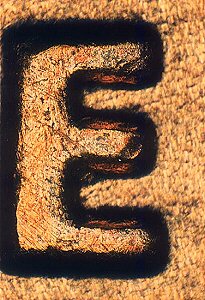
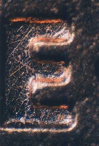
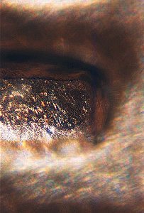
A primary use of such a microscope is the study of highly
polished and chemically etched metal surfaces. Another common use
is the inspection of flat surfaces with fine detail such as
microchips. The photographs below are of the E in the motto "E
Pluribus Unum" on a Lincoln Penny. Including the
shadow, the E is 0.3mm high. The oblique illumination photographs
used a Nicholas Illuminator from a stereomicroscope as the light
source. This provides a parallel beam of light much larger than
the field of view, and the only control (besides the lamp
position) varies the voltage to control the light intensity. As
can be seen in the photographs, the diaphragms in the axial
illuminator provide control over the depth of field and superior
glare control compared to the Nicholas illuminator, especially
for high power objectives with scant working distance. These are
important for the intended uses of the instrument.
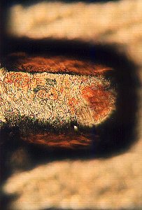
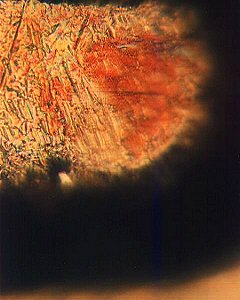
Axial illumination using 8X, 20X and 40X
objectives.
Common biological subjects are generally not especially flat,
and the lower powers are by far the most frequently used for
opaque subjects. The Nicholas Illuminator is only one of many
possible oblique light sources where the working distance allows,
and the choice can have a dramatic impact on the view. Ordinary
desk lamps can be used with either frosted or clear bulbs, the
Mini-Mag flashlights are focusable, and can also be used without
the reflector for "raw" lighting. These different
lighting techniques complement each other, their versatility is
surprising, and the necessary equipment obtained at little cost.
Oblique illumination using 8X, 20X and 40X
objectives.
Comments to the author John Wojtowicz welcomed.
An article on basic incident illumination using a compound microscope is in the Micscape
Library
© Microscopy UK or their
contributors.
Published in the April 1999
edition of Micscape Magazine.
Please report any Web problems
or offer general comments to the Micscape Editor,
via the contact on current Micscape Index.
Micscape is the on-line monthly
magazine of the Microscopy UK web
site at Microscopy-UK
WIDTH=1
© Onview.net Ltd, Microscopy-UK, and all contributors 1995 onwards. All rights
reserved. Main site is at www.microscopy-uk.org.uk with full mirror at www.microscopy-uk.net.