SOME
OF THE TYPES OF MICROFOSSILS AND NANNOFOSSILS FOUND IN THE DORSET
MESOZOIC SEDIMENTS.
BY
KEITH W. ABINERI, UK.
42
West Borough, Wimborne, Dorset BH21 1NQ, UK
Tel. 01202 885547
Introduction.
The numbers of genera and species of microfossils and
nannofossils, which have been detected in the Mesozoic sediments
along the Dorset coast, are varied and complex. Furthermore
classification can be very difficult. The sheer numbers of
individual microorganisms on 1 cm2 of the area
of each peel shows that many of these sediments are largely
composed of actual biogenic material. These include crinoidal
limestone, Kimmeridgian shales, clays and limestone bands,
Purbeck micrite1 and clay beds, Cretaceous chalk and
marl2 layers etc.
Of course individual examination of separated microfossils and
nannofossils can be undertaken by crushing, sieving, decanting
and treatment with various chemicals, of the original rocks.
These procedures are certainly very useful for the classification
of many microfossils. However the cellulose lacquer peel
technique, discussed here, gives an overall picture of the
structure of the various sediments. This applies also to
cellulose acetate peels and accurately prepared thin sections.
The six accompanied illustrations, Figures (1) to (6), show a
further selection of cellulose lacquer rock peels. These relate
to some different types of microfossils and nannofossils. However
(1) shows an image of an Upper Chalk foraminifera on an unstained
lacquer peel, using darkfield illumination with XPL. This method
of lighting records the fine structure of the chamber walls.
The variety of microfossils and nannofossils.
There is little doubt that the microfossils and nannofossils,
present in rocks and sediments, are the largest part of the
geological record, in terms of variety and numbers of individual
past forms of life. Their study leads to increasing understanding
of the Earth's history, past climatic changes, past geographical
and geological developments (from plate tectonics and ocean floor
spreading), the dating of rock strata, the search for valuable
mineral deposits (petroleum exploration) and also a large part of
the history of life on our planet.
Furthermore sedimentary rocks contain many
microscopic fragments of larger plants and animals, which are
worthy of our attention and help to build a general picture of
ancient environments (e.g. Mesozoic conifer forests).
Those amongst us who are privileged to observe Nature with an
optical microscope, or an electron microscope, can become fully
aware of the exquisite complexity of our surroundings. This
applies to the study of micropalaeontology, in particular,
because the active use of the imagination is required, as well as
natural science and accurate observation to build pictures of the
remote past ages. Obviously there are many problems and
uncertainties to challenge our ideas and theories, and encourage
new thinking. This is an age of developing natural science with
the aid of rapidly advancing technology and new methods of
processing information.
It is hoped that the following illustrations and notes will be of
interest to those who wish to observe some of these microscopic
objects and features with their own microscopes. The preparation
of the cellulose lacquer rock peels does require some practical
skill, which can be acquired by "trial and error"
methods. To obtain the best results the softer layered rocks
should be sampled. The type of peel can be assessed usually with
XPL to show the material which has been removed from the rock
surface. Likewise XPL can indicate which images on the peel are
replicas, and produce no optical figures.
Various types of nannofossils and microfossils. i.e.
Figures (1) to (6).
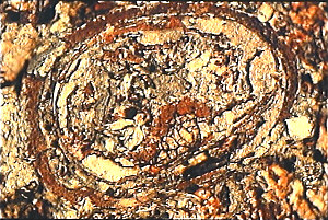
Figure (1).
This is a special dark field image of a foraminifera from
the Cretaceous, Upper Chalk, Actinocamax Quadratus Zone.
Map Reference : SY.851.802. West of Arish Mell on the
cliff, Dorset coast. Objective N.A. 0.65. Total field
area of the image = circa 170 microns x 140 microns.
|
 |
This image shows darkfield
illumination combined with XPL, with no replica images. The
oblique section is circa 163 microns in maximum diameter. The
walls of the interior chambers show considerable detail under
this mode of illumination. The species is believed to be Globotruncana.
Unstained Upper Chalk peels generally will give these results
with dark-field illumination and XPL, when applied to
foraminifera images.
Figure (1) required an exposure of about five
seconds with the ASA 100 film. Visual observations of the fine
structure of the interior chamber walls need a period of
"dark adaptation" by the eye.
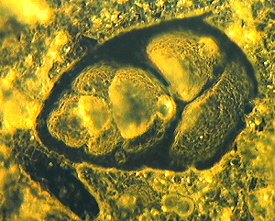
| Figure (2).
Here we have an intact Coccolithophore3
nannofossil, buried in kerogen. This image was derived
from an unstained cellulose lacquer peel, from the
Jurassic, Kimmeridge Clay, Maple Ledge Shales. Map
Reference : SY.908.790. East of Gaulter Gap, Kimmeridge
Bay, Dorset coast. Oil-immersion objective N.A. = 1.25.
Total field area of the image = circa 30 microns X 20
microns. |
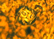 |
Figure (2) is an example of a removed thin
layer type of image. The real diameter of the Coccolithophore is
estimated at 11 microns. The surface coccoliths are circa 4.2
microns in diameter. Clearly electron microscope images are
needed for closely detailed examination. The species here is
believed to be Ellipsagelosphaera brittanica4.
Similar coccolithophore images have been identified on stained
cellulose lacquer peels, but these are usually less well defined
than on the unstained peels. However, most surprising, some of
the stained coccolithophores (and coccoliths) show a deep blue
colour due to Ferroan Calcite composition, whereas many others
are pale pink in colour suggesting very pure calcite composition.
This differential staining of coccolithophores and coccoliths
requires confirmation.
Figure (2) was obtained with brightfield illumination using one
polarizing plate above the objective.
| Figure (3).
Here we have images of Dinoflagellate5
cysts from a stained cellulose lacquer peel. The
highly magnified picture was derived from the
Jurassic, Upper Kimmeridge Clay, Freshwater Steps
Stone Band. Map Reference : SY.943.772. Freshwater
Steps, west of Hounstout Cliff, Dorset coast.
Oil-immersion objective N.A. = 1.25. The total field
area of the image = circa 100 microns X 70 microns. |
 |
Mainly the images of these microfossils on the
cellulose lacquer peels are buried in the kerogen and/or
encrusted with coccoliths or coccolithophores. The best images
are obtained for classification by the techniques of Palynology6, which involves the removal of all the mineral contents
of the sediment, using concentrated hydrofluoric acid7. This technique is used also for the isolation of
pollen grains and plant spores. Slides of the organic material
only are prepared in a Canada-balsam mounting, as a strew of
microfossils for microscopic examination. An important
paper by N.S. Ioannides, G. N. Stravrinos and C. Downie l9768 based on the Kimmeridgian microplankton from Clavell's
Hard, Dorset coast, classified some sixty species of
Dinoflagellate Cysts and came to some important conclusions about
the origins of the kerogen in the Kimmeridge Oil Shale9.
Figure (3) was obtained with brightfield illumination and PPL.
The complete diameter of these Dinoflagellate cysts range from
circa 20 to 50 microns.
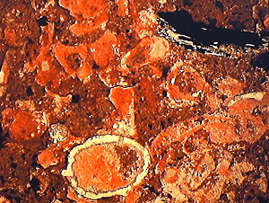
| Figure (4).
Here we have a bed of broken Ostracod10
carapace sections on a stained lacquer peel from the
Upper Jurassic, Middle Purbeck, Lulworth bed 107. Map
Reference : SZ.036.784. Durleston Bay, nr. Swanage,
Dorset coast. Objective N.A. = 0.10.
The total field area of the image = circa 2340
microns x 1920 microns. |
 |
Ostracods are microscopic Crustacean arthropods11 of great geological interest and are frequently studied
because of their widespread occurrence in various sediments. As
arthropods, living Ostracods are complex multicellular animals
with a digestive system, a central nervous system and developed
genital organs. The bodies appear unsegmented and are contained
in a calcitic carapace consisting of two valves. The microfossils
are generally found as carapaces only.
In Figure (4) the prominent Ostracod carapace has a maximum
diameter of about 813 microns. There are also fragmented
carapaces present, as well as a fragment of conifer wood,
consisting of removed fusain and replica type imagery. The red
colour on the peel is due to stained Micrite. The darker colour
may be due to Marl or clay. Lulworth bed 107 in Durleston Bay
lies just below the well-known "Cinder Bed", which
marks a temporary marine incursion, and is the accepted boundary
between the Jurassic and Cretaceous periods. The "Cinder
Bed" shows masses of marine Oyster-rich sediment but the
beds below it show numerous freshwater fauna, including
Ostracods.
Figure (4) was obtained with
brightfield illumination and one polarizing plate above the
objective.
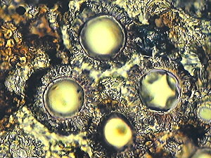
| Figure (5).
Here we have the image of a single Ostracod carapace
section on a stained cellulose lacquer peel derived
from the Upper Jurassic, Middle Purbeck, Lulworth bed
102. Map Reference : SZ.036.784. Durleston Bay, nr.
Swanage, Dorset coast. Objective N.A. = 0.25. The
total field area of the image = circa 1350 microns X
900 microns. |
 |
This Cypridea Ostracod12 carapace section has a main axial diameter of about
1210 microns. This lacustrine and brackish water type of Ostrocod
is widely distributed in the Purbeck freshwater beds. The
Cytheracea Ostracods13 are found
in marine environments. The red stained structures on Figure (5)
indicate calcite and micrite composition. Marl and clay areas are
also shown. The peel was examined with brightfield illumination
and one polarizing plate above the objective. Some fusain can be
seen on this image.
| Figure (6).
Upper Chalk sediments contain numbers of microfossils,
nannofossils, calcisphereres, foraminifera, fragments of
larger life forms etc. However here we have an
unidentified microfossil with a flask or bottle-like
appearance on a very detailed stained cellulose lacquer
peel. This picture is from the Cretaceous, Upper Chalk,
Actinocamax Quadratus Zone. Map Reference : SY.851.802.
West of Arish Mell on the cliff, Dorset coast. Objective
N.A. = 0.65. Total field area of the image = circa 250
microns X 170 microns. |
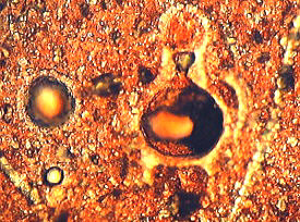 |
The differential staining of the minute
nannofossils on this cellulose lacquer peel are accentuated by
the brightfield PPL illumination. Apart from the flask or
bottle-like microfossil image, Figure (6) shows calcispheres and
numerous nannofossils, as well as probable sponge fragments. This
unique flask-shaped object either represents an unknown species
(perhaps a type of Calpionellid14)
or may represent the cell fission of an unkown microorganism.
Notes and References.
1. Micrite : micro-crystalline calcite
(grain size less than 10 microns).
2. Marl : a calcareous mudstone.
3. Coccolithophores : coccospheres, a variety of marine algal
Phytoplankton made of coccolith calcareous plates.
4. Ellipsagelosphaera brittanica : a species of
coccolithophore which occurs in the Kimmeridge Clay mainly as
dispersed coccoliths.
5. Dinoflagellate cysts : a family of organic microfossil plant
cysts best separated by palynological techniques by treating the
rock with concentrated hydrofluoric acid and some concentrated
nitric acid. Marine Phytoplankton.
6. Palynology : The separation technique used in 5 above, and
also for the isolation of pollen grains and other land based
plant spores.
7. Hydrofluoric Acid a powerful mineral acid which will dissolve
all silicate and other mineral rocks.
8. N. S. Ioannides, G.N. Stavrinos and C. Downie, 1976.
"Kimmeridgian microplankton from Clavell's Hard, Dorset,
England." This important paper reports and describes some
sixty species of Dinoflagellate Cysts from the Kimmeridge
Blackstone (oil shale). However from the high numbers of
Terrigenous palynomorphs (land based pollen and plant spores) in
the oil shale layers, the authors deduced that much of the
Kerogen was derived from land widespread swamp floras, due to a
slight rise in sea level, and flooding into the Kimmeridgian sea.
A close correlation was observed between the quantity of Kerogen
and the proportion of terrestrial palynomorphs.
9. Kirmmeridge Oil Shale : Variable amounts of these
deposits, rich in Kerogen, point to fluctuations in rainfall,
flooding and sea levels. Kimmeridge Clay seems to be one of the
source rocks for North Sea Petroleum.
10. Ostracod Carapaces : These occur in vast numbers in the
Purbeck beds. Marine, brackish and freshwater species of these
Crustaceans have occurred in geological times.
11. Arthropods : Animals with Jointed legs. A large phylum
including classes Crustacea, Arachnida, Insecta, Myriapoda and
Trilobita.
12. Cypridea Ostracod : A species of ostracod found in Lacustrine
and brackish water environments.
13. Cytheracea Ostracod : A species of ostracod found in marine
water environments.
14. Calpionellid : A flask-shaped microfossil.
Editor's note: Some of the
quality of the author's original 35mm slides is lost in the
scanned and compressed web images. Comments to the author are
welcomed, who can be contacted at the above address or comments
can be passed on via the Micscape Editor, see contact on magazine
index.
Other articles in this series can be accessed
in the Micscape on-line library by typing in the author's surname
'Abineri' in the Library search engine (link below).
Prepared for the Web by David Walker
© Microscopy UK or their
contributors.
Published in the April 1999
edition of Micscape Magazine.
Article at
http://www.microscopy-uk.net/mag/artapr99/kamast3.html
Please report any Web problems
or offer general comments to the Micscape Editor,
via the contact on current Micscape Index.
Micscape is the on-line monthly
magazine of the Microscopy UK web
site at Microscopy-UK
WIDTH=1
© Onview.net Ltd, Microscopy-UK, and all contributors 1995 onwards. All rights
reserved. Main site is at www.microscopy-uk.org.uk with full mirror at www.microscopy-uk.net.