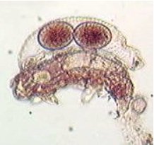
A sample of water from Lochaber, Scotland collected by Alastair Bruce was received on the 1st. August 2000. This was examined on receipt and part preserved in formalin, my usual practice. On subsequent days temporary mounts were made of live material sealed with petroleum jelly and searched for desmids, my main interest. A mount was made on the 8th. August and examined. On the 9th the slide was examined again and a tardigrade was found moulting, see photomicrographs. It was still alive and squirming with the shed skin still attached 24 hours later.
 |
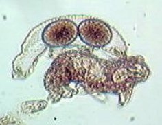 |
The shed skin in which two eggs have been deposited
can be seen above the tardigrade in both photo's. The eggs are circular
in end view. The skin has regularly spaced rows of papillae on the surface
throughout its length, the papillae can just be seen in the photo's and
in these side views they give a crenate appearance along the back. As the
shed skin is curved, its length was difficult to measure, however it measures
189 µm from head to tail, allowing for the curve it must be about
200 µm.
The general appearance of the tardigrade and the sculpturing on the skin surface matches the description of Hypsibius (Isohypsibius) annulatus (Murray): Marcus 1929.
Tardigrade, Latin tardus = sluggish. gradus
= a walk.
References;
Clegg, John FRESHWATER LIFE Revised edition 1974.
F.Warne & Co.
Morgan C.I & King P.E. BRITISH TARDIGRADES
1976, The Linnean Society of London.
Comments on the above by e-mail are welcomed to Bill Ells.
William Ells.