Epizoics on a mosquito larva

Michael Reese Much

Bethlehem, Pennsylvania, USA


Click each image to view a larger version


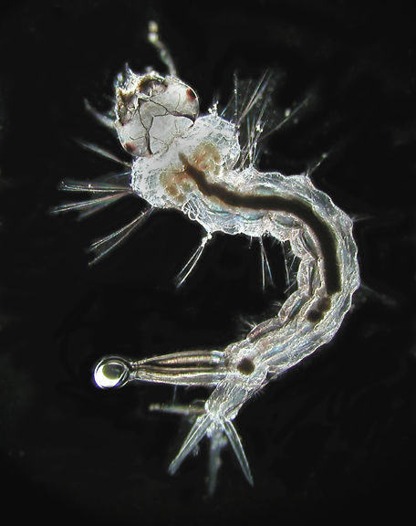
One of the more fascinating subjects for the microscopists are mosquito larvae—commonly known as “wrigglers.” These fairly large specimens—8 to 10 millimeters in length—barely fit into the field of view of a 4X objective. The image to the left is at 40X. At it’s tail end the wriggler has a breathing tube (there is a bubble on the tip of this specimen's tube) and around its anus a cluster of feather-like fins.
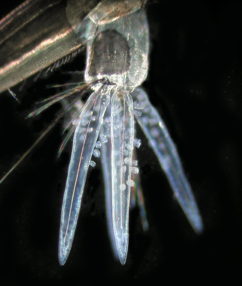
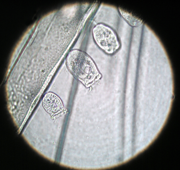


A recent water sample from a marsh contained a single wriggler. Upon examining it I discovered the tail fins were covered with epizoics (likely to include stalked peritrichs and others such as rotifers as some were observed to swim away and return). I returned to the site and collected more wrigglers to obtain better specimens. I found that some wrigglers had as many as 80 examples clinging to the fins.


40x


The epizoics appear not to harm the larva but seem to feed on minute particles passing through the wrigglers gut—and a wriggler is almost all gut. The dark mass running through its body is fecal matter. I attempted to get “cleaner” specimens by progressive changes of water, but the larvae are very efficient feeders.

The epizoics latch on to the fins with their stalks and occasionally swim away and then return. Wrigglers prefer to float head down in the water with their breathing tube poking through the surface of the water. When alarmed they thrash about quite violently.


100x


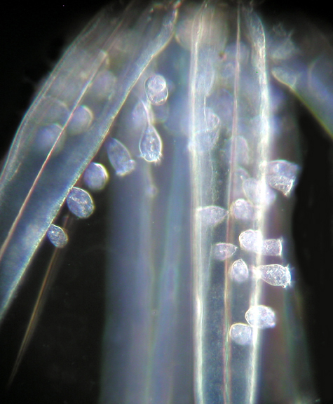
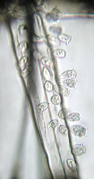
If you see how severe the thrashing motion of a swimming wriggler is you can appreciate how firmly the epizoics are able to grip the fins of the wriggler. The view to the left is at 400X.

The relationship of the epizoics and mosquito larvae are not one of a parasite and host in the truest sense. It doesn’t seem that the they are harming the wriggler other than being an annoyance. It is more like the relationship of remoras to sharks—the remoras adhere to the sharks to gather scraps of food from the sharks kills. The epizoics seem to be feeding off the wrigglers' wastes.

It may be that the wrigglers get some payback from their uninvited guests. I noticed some larvae specimens occassionally preened their hindquarters. No doubt they are eating the epizoics they nibble off. Talk about a vicious cycle!
400x


A few comments about specimen preparation:
I mentioned previously that I put the specimens through progressive changes of water. This was done primarily to reduce background clutter for the photography. I tried staining by putting the wrigglers in water containing methylene blue, carmine and crystal violet for five minutes before making the slide but saw little effect with these stains. The crystal violet appeared to stain some food at the beginning of the gut. I put the water drop on the slide, drew off most of the water with a toilet paper wick, replacing the water with methyl cellulose. 22mm cover glasses did not harm the wriggler but 24X40mm slips crushed them (maybe more methyl cellulose would float the slip better).


400X phase lighting.


I’ve been buying stuff from a retired pathologist and got several thousand of these 24X40 slips for $20. I got a Spencer microtome for $50. If anyone needs an automatic slide processor(dehydrating, staining) he’s got one for $100.

I am shooting with an Olympus C-4040 digital camera on an Omano trinocular microscope modified with a Wild achromatic aplanatic variable phase condenser. I am in the process of upgrading my objectives. If anyone knows of any objectives with iris diaphragms in them that are available I would appreciate the lead.

1000X oil immersion.

All comments to the author Michael Much are welcomed.  

Acknowledgement: Thank you to the readers who provided feedback on an earlier version of this article and who provided help on the identification of the epizoics.


Microscopy UK Front Page
Micscape Magazine
Article Library


© Microscopy UK or their contributors.

Published in the August 2004 edition of Micscape.
Please report any Web problems or offer general comments to the Micscape Editor.
Micscape is the on-line monthly magazine of the Microscopy UK web
site at Microscopy-UK  


© Onview.net Ltd, Microscopy-UK, and all contributors 1995 onwards. All rights reserved. Main site is at www.microscopy-uk.org.uk with full mirror at www.microscopy-uk.net.