 |
APTENIA
CORDIFOLIA a short illustrated monograph |
|
|
| |
APTENIA
CORDIFOLIA a short illustrated monograph |
|
|
|
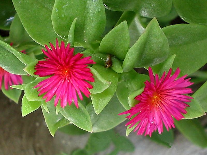
INTRODUCTION My very small tropical garden is especially composed of leafy plants (plants which seldom flower but which show very attractive foliage forms and colors, like Epipremnum , Syngonium , Chlorophytum, Codiaeum, or "the Water wood" (Dracaena fragans), Tradescantia (and many others) and by small flowering plants (Portulaca (2 sps), Aptenia , Hybiscus, "alegrias", Kalanchoe). These last are, moreover, plants which can reproduce from cuttings, of very fast growth and of easy dissemination. Portulaca oleracea , Aptenia, and Syngonium podophyllum, Epipremnum aureum or Chlorophytum comosum too, are very attractive to be used in flowerpots with hanging foliage which are very much used to give greenery and freshness inside and outside of our houses. Using a search engine you can find pictures of all of these on many garden or botanical sites on the Internet. Few of them (even the most characteristic and abundant in the local gardens) are autochthonous plants. Aptenia cordifolia (Lf) Schwante, which I present here, is a modest trailing plant, of waxy appearance, soft consistency and with small red flowers. It was transferred to Europe from an area near Johannesburg and subsequently dispersed in the Americas , starting with an importation made to the United States in 1970 from Israel . It is very common in Mexico , and I used it in Durango to obtain epithelium samples (which have a very interesting morphology, and are very easy to peel from the leaf underside) to be used in my work on the mounting media. In Cancún small cuttings planted in sunny places, without any greater precaution, produce dense plants full of flowers in a few weeks. If one doesn’t wish that it becomes a plague, its growth must be carefully controlled, because it outgrows any other plant which appears in its way. Its best destiny is to be cultivated in flower-pots.
The title picture shows the foliage and the flowers of a young plant. We will see the more important macro and microscopic features of this plant in two sections: 1) vegetative organs and 2) reproductive organs.
VEGETATIVE ORGANS
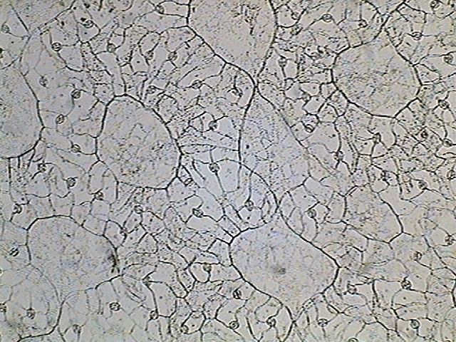
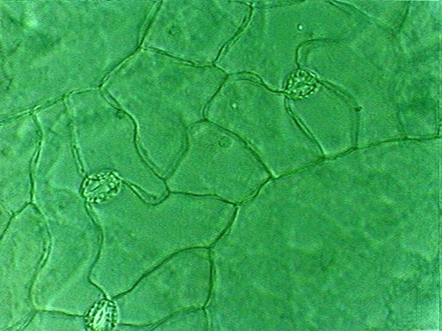
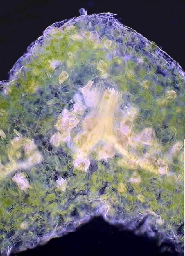
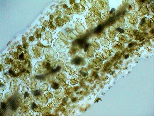
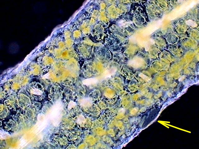
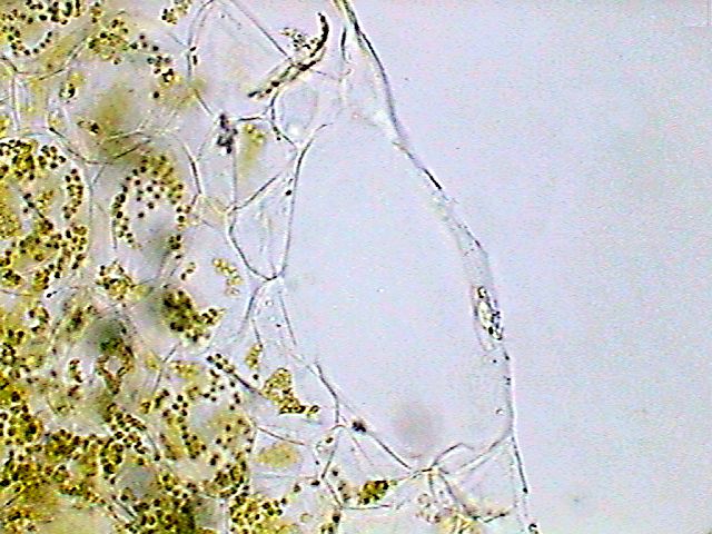
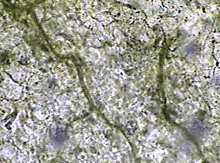
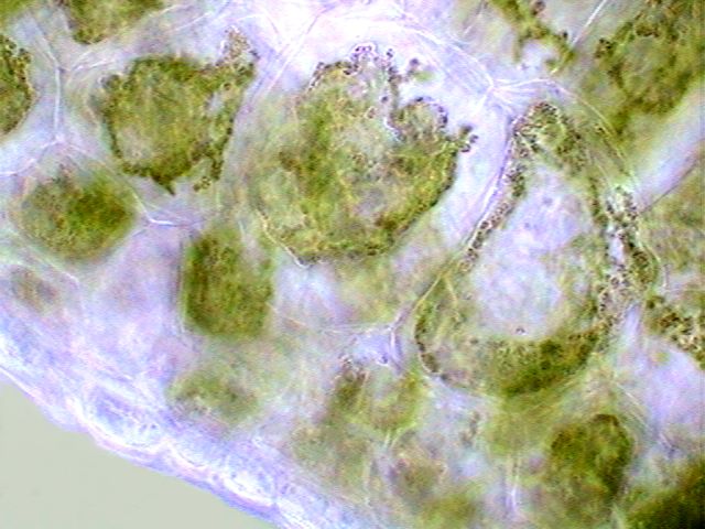
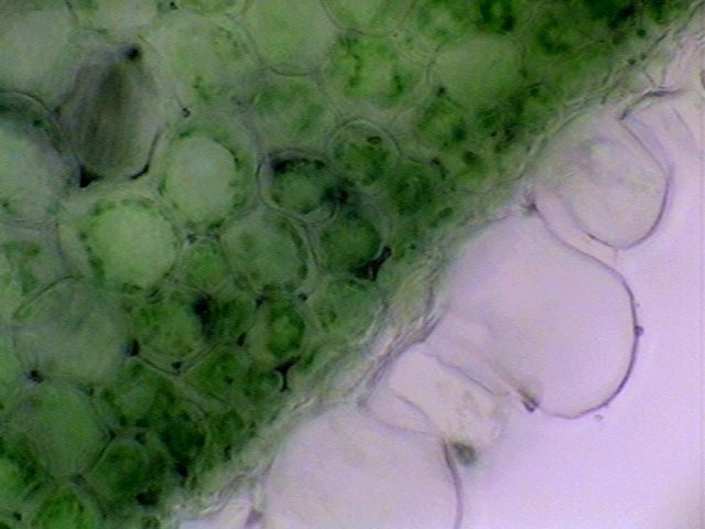
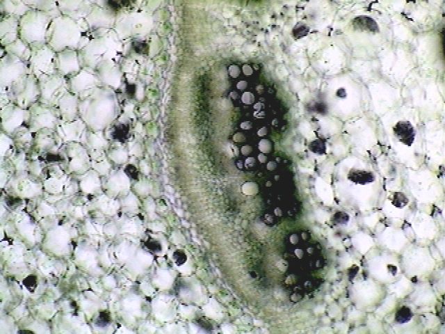
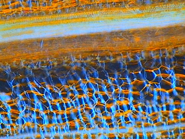
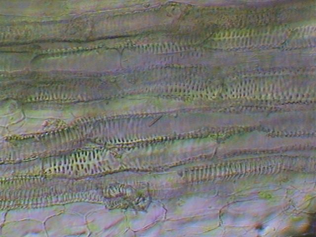
THE LEAF. The following pictures show the epithelium of the LEAF at three different powers. Leaves are hypostomates that is to say that they have the stomata in the lower or ventral face of the leaf. As one can see very well in the picture captured with the 100x objective, the stomata are simple, both guard cells being surrounded by normal cells of the epithelium. On the contrary, composed stomata, like those of Tradescantia, are surrounded by additional cells different from the normal epithelial cells, although they also do not have chlorophyll. (See a picture, here) There is a special detail which I saw in the epithelium of no other plant, either those on which I made a direct study, or in the images that I have seen. They are those which I call lacunar cells,
very large, much thickened, with an internal enormous vacuole and a
thin peripheral cytoplasm, which has a very large nucleus, generally on
the external layer of the cytoplasm. These cells form a very
characteristic network, as can be appreciated in the surface image
taken with the 10x objective.
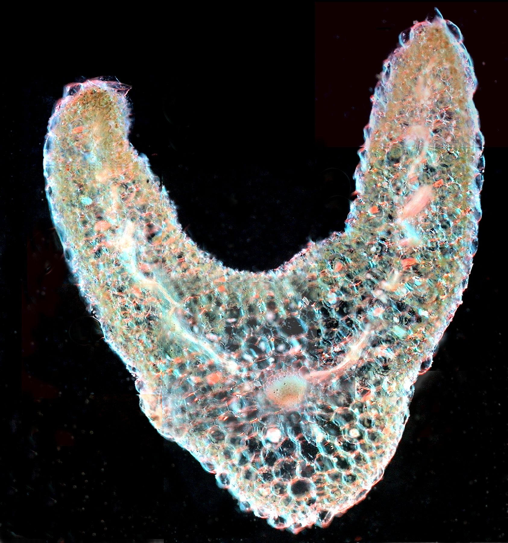
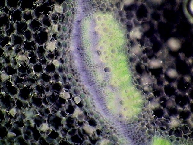
The foliar epithelium is the only part of this plant which permits easy research. Leaves and young stems have a very soft consistency and a loose structure, in which the cells appear to float in a mucilaginous medium. To obtain clean sections, which also cannot be too thin, the edges of the mesotome must be much sharpened. For better success it is well to let materials desiccate for 2 or 3 hours. One obtains a much firmer texture. Another particular characteristic of the leaves is that at the start I could not identify a vein or a definite network of veins. Like all the leaves, those of Aptenia have, as it can be seen in the first picture, the shape of a butterfly with a central gutter which makes us suppose the existence of a main vein. However as soon as I carried out one transverse section of the leaf crossing the "gutter", I have been able to see only one continuous blade which, even under the objective, lost its 'butterfly' shape very quickly. Some randomly oriented vessels were identified where the central vein of other leaves are clearly seen. Compare with other leaf sections in my previous papers. One can find a central vein only in the posterior third
of the leaf but with a weak contrast which makes difficult its
identification.
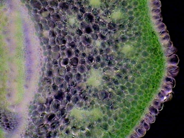
And the vessels, which moreover are numerous, are cut in
small sections, as if they had a very sinuous path, without a well
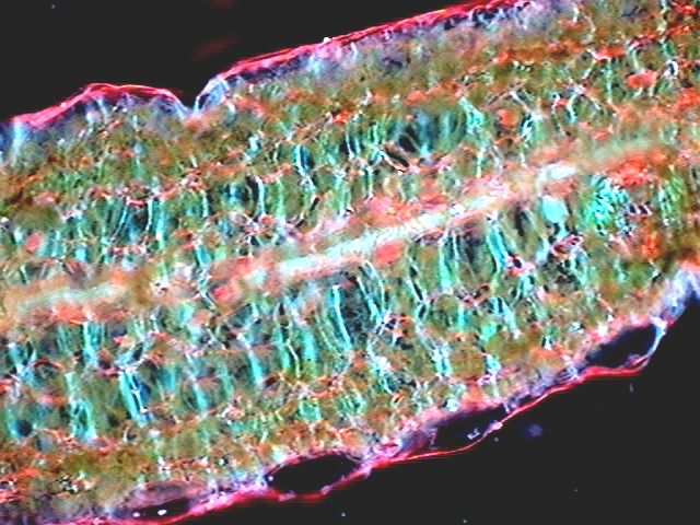
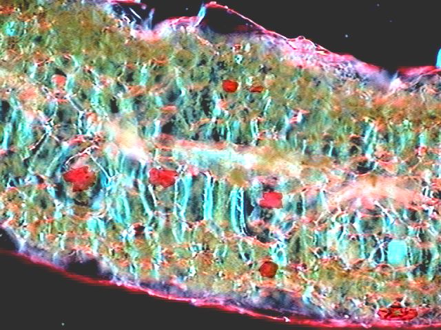
defined direction. In the following stitched pictures (figs. 7 and 8)
the vessels are the central, longitudinal, dense structures (dark in
the brightfield image; and bright in the
darkfield one). The small whitish spots distributed in the parenchyma
are clusters of raphides.
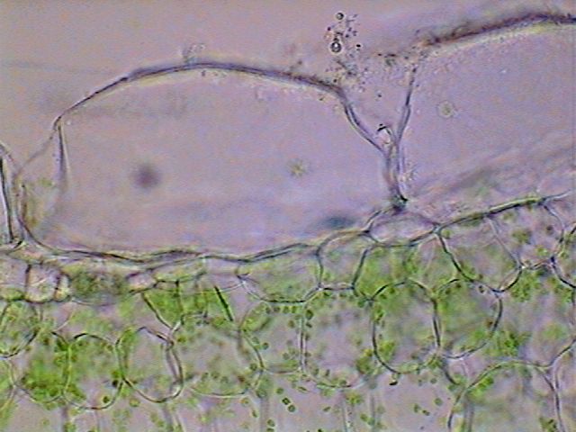
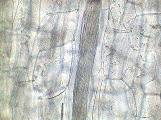
In the sections of
the leaf blade one can define the two layers of epithelium (higher and
lower). In the lower epithelium the large and long lacunar cells are
easily differentiable.
An image of a lacunar cell with a higher power is shown in the following brightfield picture (fig 11).
Searching for the "veins". In order to be able to see the distribution of veins in the leaf mesophyll I used the following technique. a) Washing out the chlorophyl .- 2 leaves were boiled in water for 10 minutes in order to well soften the cellular walls, and they were then boiled 20 min. (80ºC) in 96% alcohol in order to eliminate chlorophyll. The process bleaches leaves completely. b) Clarification.-
To see
through them they could have been impregnated with glycerin (IR=1.47).
As I did not have it at the time, I used castor oil (IR=1.48) that it
is also miscible with alcohol 96. After 3 hours of contact, the leaves
are transparent. One of them was mounted between slide and coverslip,
but as it was curly I pressed it with 4 weights of 15g for 14 hours.
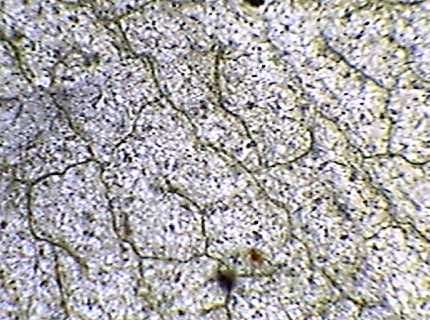
c) Seeing through the leaves shows precisely what I suspected. In the greatest part of the leaf the vessels describe a sinuous form with the result that a right cut shows them like separate segments. And they form a network (which, curiously, one does not see as completely connected), which covers all the leaf by forming a fine mesh, but not a main vein as in most leaves. d)
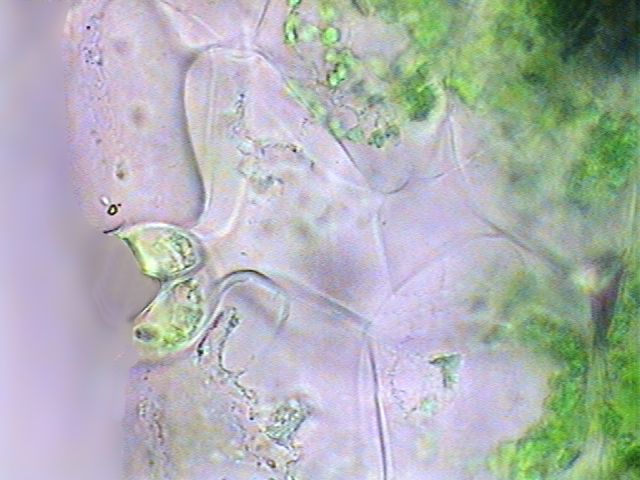
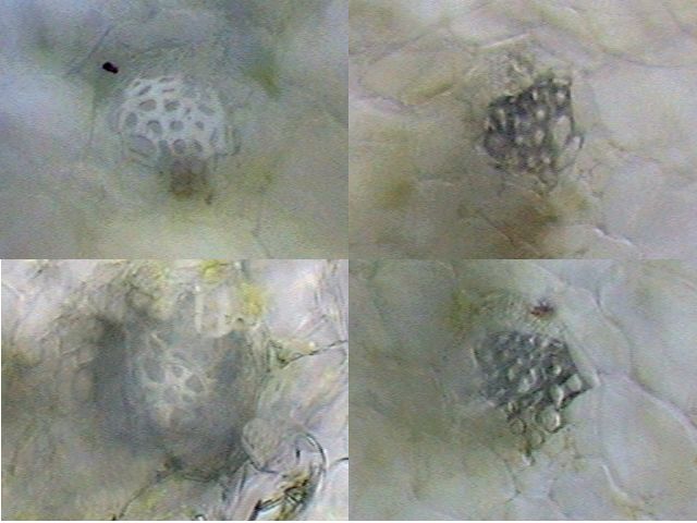
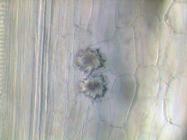
They are less easy to see the sections of the stomata,
and one must look between the cells of the lower epithelium of the
leaf. But in the following figure, one sees the two guard cells charged
with chlorophyll and under them a pear-shaped air cavity, the stomatic cavity (fig.16).
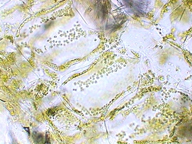
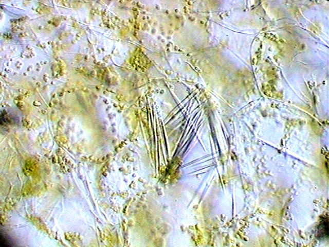
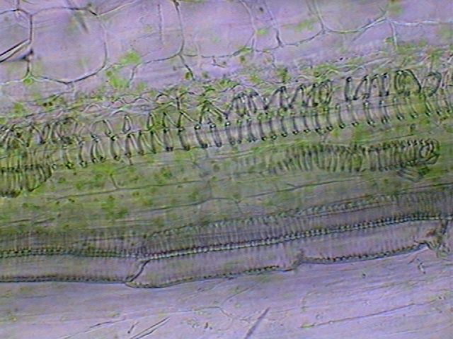
There is a sub-epithelial layer of 2 or 3 small cells, globose, charged with chlorophyll (very visible in darkfield) and below, or intermingled with them, a layer with large rectangular cells, also with chlorophyll, which appear to represent the palisade epithelium of more conventional leaves (fig. 17 and 19). The two or three following layers represent a
mesophyll with globose cells, between which there are an abundance of
idioblasts with acicular raphides of calcium oxalate (fig.18).
FOLIAR
PETIOLE. The petiole of the leaf is more difficult to interpret.
It shares the same loose structure of the leaf, although it has a more
defined indicator of a principal vein (fig. 20). The epithelium now
takes the special form which we will study in detail in the stem. The
cross sections must be observed quickly to prevent them completely
losing their structure in a few seconds.
Pictures were taken using a Rheinberg filter with crimson
center and greenish outer ring. Its structure corresponds obviously to
that of a dicotyledon. From the outside towards the interior one can
initially describe an epithelium which is very special in many ways.
Contrary to what the epitheliums of other plants that I examined are,
which generally show its protective function by the thickened external
cuticle, often covered of wax, and by the small and thin cells which
conform them, this epithelium of Aptenia is formed by long and high
cells, of round external profile with the shape of a cushion (with
rectangular plant, which in the transverse section are seen like
transparent globules of fine cellular wall) (figs.22 and 23).
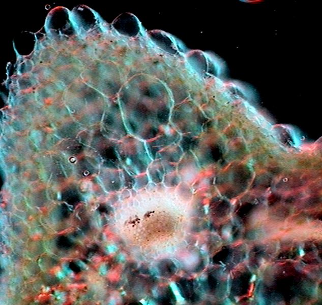
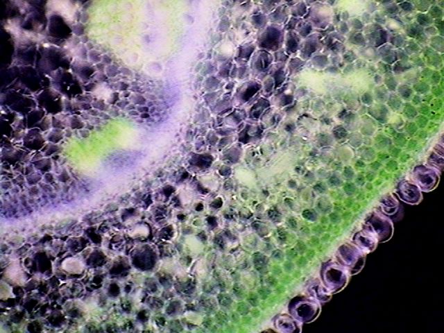
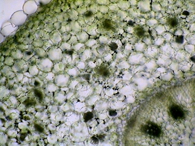
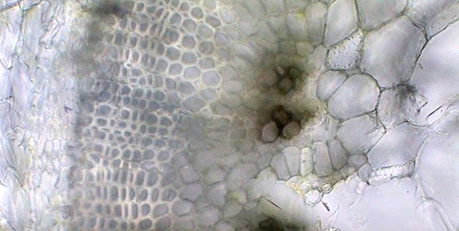
To recognize its true form it is necessary to examine the longitudinal section of stem which we present in fig. 29. Under the epithelium there is a sub-epithelium with 2 or 3 layers of cells with chloroplasts, and then we see a cortical parenchyma. Dispersed inside the
cortical parenchyma one finds several small packages with some fibers,
and few small vessels of xylem (2, even 4 - Fig. 24), in addition to
many crystals of calcium oxalate. The vascular packages in the cortical
parenchyma are atypical in a dicotyledon, but they are clearly
recognizable (but difficult to photograph - they are very dense and
obscure). They generally have two or three woody vessels of very small
diameter.
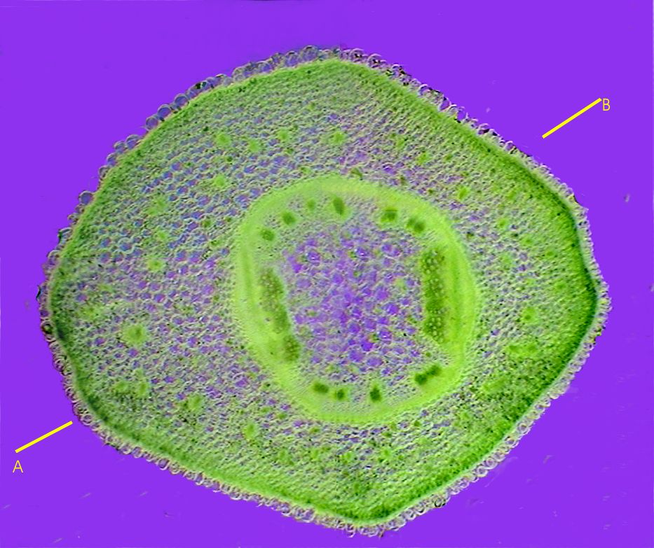
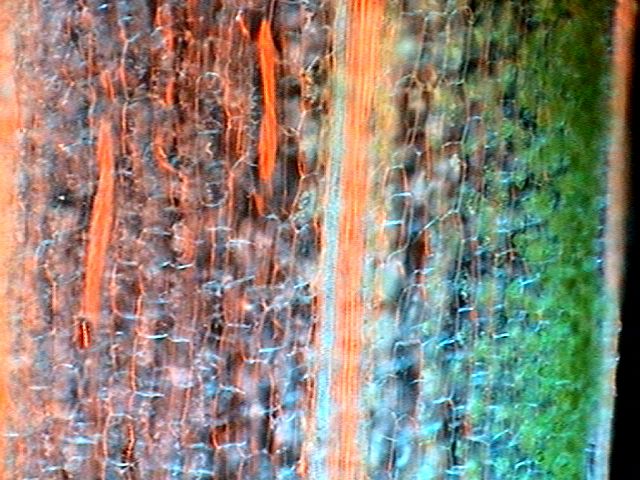
The cortex is limited internally by a layer of cambium (refringent yellowish lines, in figs. 21 and 22) which surrounds the central cylinder within the limit of which one finds the principal vascular packages, with a very specific design. At the two sides of the central cylinder one finds two vascular bundles provided with phloem, xylem and fibers, in definite layers. (figs. 27 and 28)
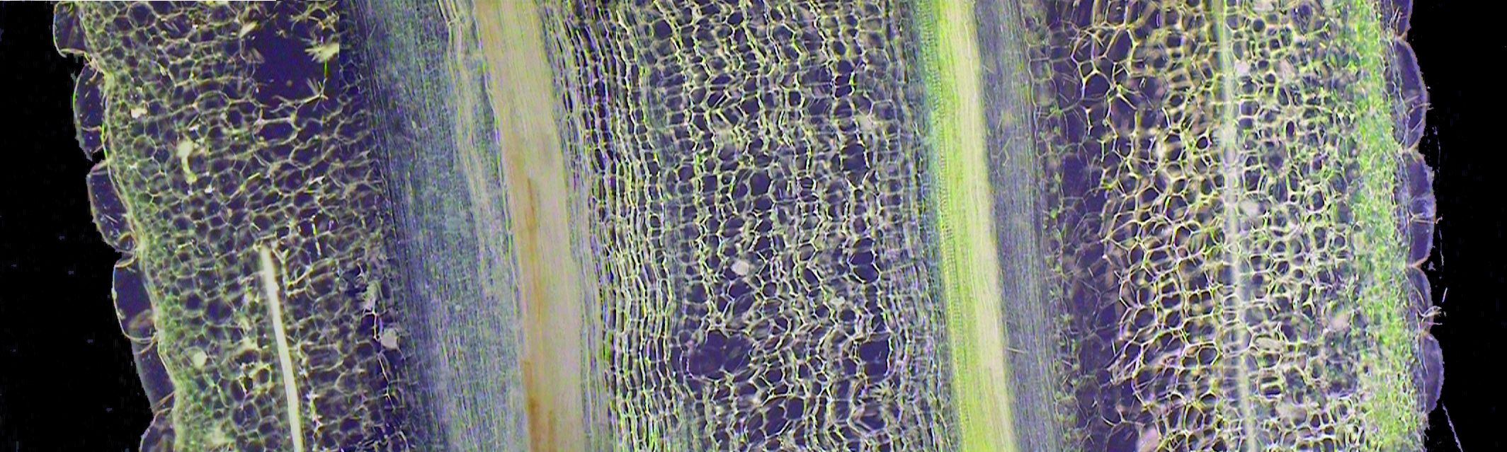
Moreover they are between these large packages, located in equidistant positions, 5 small sized bundles in one semicircle, and other 5 in the opposed one. (figs.- 21) Pith parenchyma has large globular cells which also show drusses. Line A-B drawn through the transverse section in fig. 21 shows a transect which could provide a longitudinal section similar to that really made. To show the most important anatomical details we will use a rebuilding of the section prepared with 4 individual pictures, taken with the X10 objective (fig. 29).
A t the two sides of the section we confirms the globose and aqueous nature of the epithelial cells which now shows their long vertical dimension, at least 3 times larger than the transverse diameter. With the chosen power one can only roughly define the characteristics of various tissues although is quite visible in its topographic distribution and its nature. But in the stitched band they are quite visible the details which we have described in the coss section: the epithelium followed inside by a chlorophyllian sub-epithelium, the cortex, in which one appreciates the chalky lines of cortical fiber beams (complete at right image, incomplete on the left, surely because it was cut obliquely), and the parenchymatic cells themselves with loose and aqueous aspect. Details below enable us to better describe their histology. Pictures 30 to 33 show aspects of the cortex
and the following ones (34 and 35) details of the vessels
of a main bundle :
A sector of a cross section that shows from the pith
parenchyma to the phloem is included here.
In the next part we will see the structure of
the reproductive bodies.
|
Please report any Web problems or offer general comments to the Micscape Editor.
Micscape is the on-line monthly
magazine of the Microscopy UK web
site at Microscopy-UK