|
|
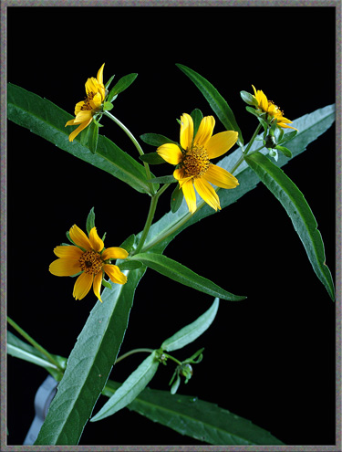
A
Close-up View of Four Beggar-Ticks: (Bidens frondosus, Bidens vulgatus, Bidens
tripartitus & Bidens cernuus) |
|
|
A
Close-up View of Four Beggar-Ticks: (Bidens frondosus, Bidens vulgatus, Bidens
tripartitus & Bidens cernuus) |
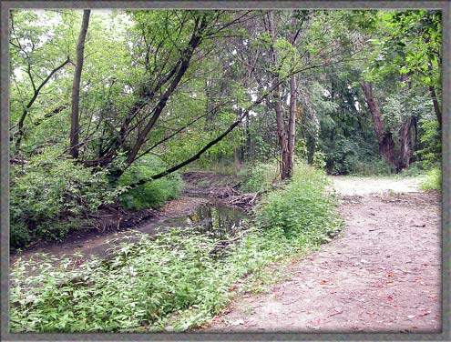
Late
in the summer, several interesting species of wildflowers called “Beggar-Ticks” bloom along the edge
of a stream near my home. All are members of the Aster family,
whose flowers normally consist of both inner “disk” florets and outer
“ray” florets. The beggar-ticks however, are somewhat unique in
that some have ray florets, some have a variable number of stunted ray
florets, and some have only disk florets!
The strange name of the group, “Beggar-Ticks”, is related to the
double-barbed seed which easily sticks to the pants of beggars, (and
others), who come into close contact with the plant in the fall.
In fact the genus name Bidens
comes from the Latin bis,
meaning "twice", and dens,
meaning "tooth", referring to the two barbs on each seed.
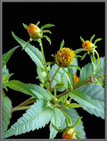
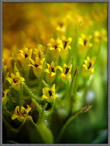
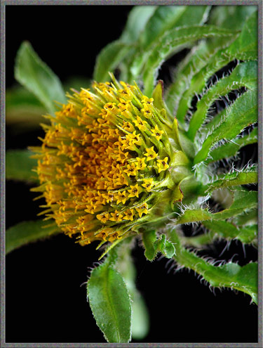
Devil’s Beggar-Ticks - Bidens frondosus
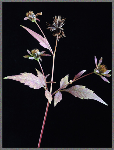
Along the edge of the previously mentioned stream, this is by far the
most common Bidens species. Most plants grow to about 80 cm in
height and have many yellow-orange blooms. A typical example is
shown as the first image in the article. Each flower-head tends
to be from 1.0 to 1.5 centimetres in diameter.
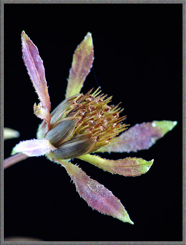
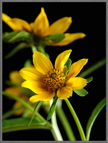
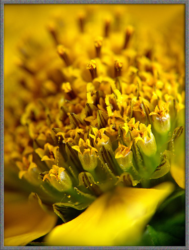
A closer look at a bloom reveals the disk-like head containing many
individual flowers, and the surrounding tiny leaflets called “phyllaries”. Devil’s
Beggar-Ticks have ten or fewer of these phyllaries.
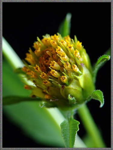
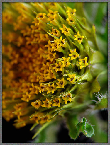
Each of the orange disk flowers has five petals. Notice that the
flower closest to the viewer is on the end of a short rod-like stalk
which grows out of a pale green structure. This is the ovary containing the ovule that will
develop eventually into a seed. The two pale green spikes framing
the flower are the immature spiny bristles that will eventually help
carry the seed to a new location stuck to the beggar’s pants!
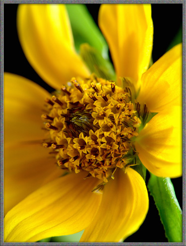
The image below shows the flowers from above, with their almost black stamens and pistils.
Surrounding the outer edge of each flower-head is a double row of phyllaries, (modified leaves), that
are quite different than the ones mentioned earlier. These are
broader, much shorter, and finely striped in purplish-black.
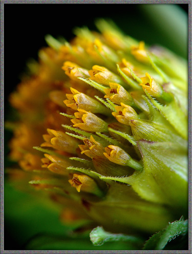
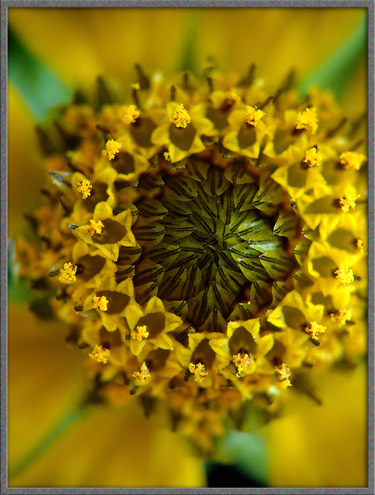
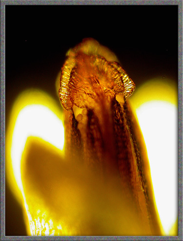
Under the microscope it is possible to see details that cannot be
resolved by macrophotography. The following two images show the
developing backward pointing spikes on the immature bristles attached
to the ovary.
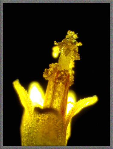
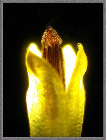
A photomicrograph of a single flower reveals the component parts.
Projecting out from the tiny petals is a large diameter tube formed by
the five fused stamens, three of which can be distinguished in the
photomicrograph. At the top of each is an enlargement, the anther (male, pollen producing
organ). Projecting out of this larger tube is the narrower style, holding at its tip, the stigma (female, pollen accepting
organ). In the image, both anthers and stigma are encrusted with
pollen.
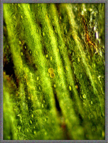
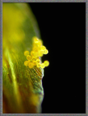
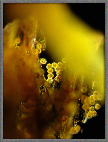
Below, on the left, two yellow spiked pollen grains can be seen
adhering to the surface of one of the striped phyllaries mentioned
earlier. The image on the right shows a group of pollen grains
attached to the edge of a phyllary.
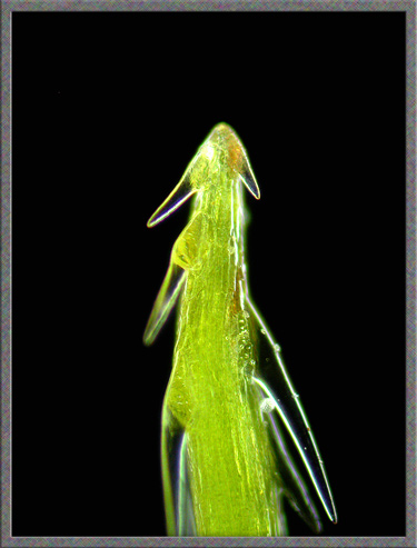
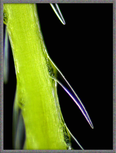
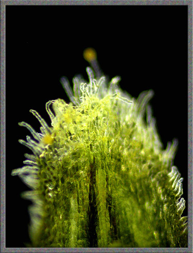
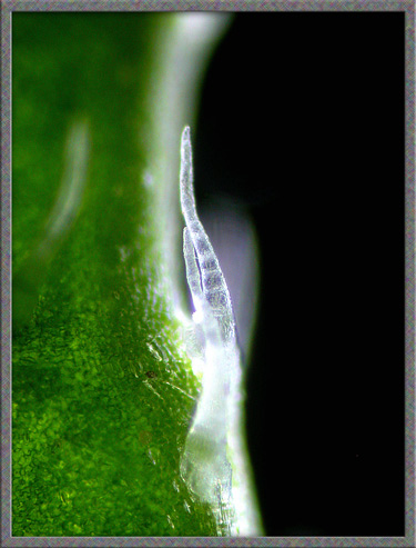
The tip of one of these phyllaries is shown on the left. Note
that it is covered with tiny transparent, hair-like projections.
The higher magnification image of one of these hairs reveals that it is
segmented.
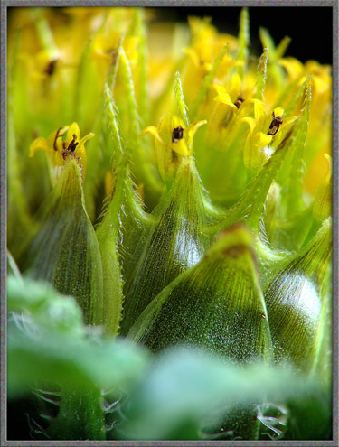
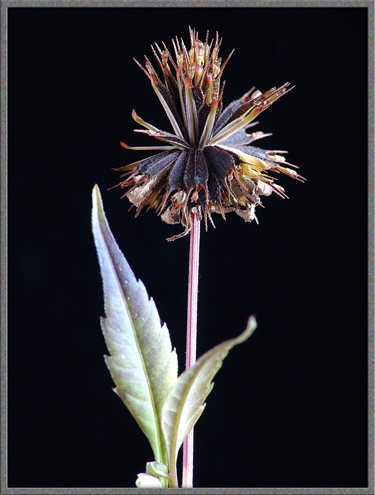
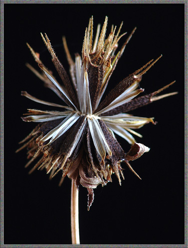
In early fall, the entire plant takes on an attractive coppery
colour. The image below left shows three younger flower-heads and
an older one that has opened up into a late stage seed-head. On
the right is a higher magnification image of the former. The
photograph clearly shows the outer ring of leaflet-like phyllaries, the
inner rings of striped phyllaries, and the maturing seeds.
In the last stage of development, the now mature fruit, seeds called
achenes, open out into an almost globular shape. This
configuration puts the spiked barbs on the outer surface of an
imaginary sphere, and provides them maximum access to any passing
ambulatory transporter.
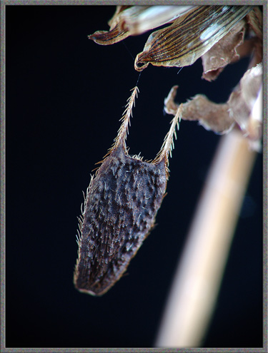
The seed of a Beggar-Tick may not be beautiful, but I can attest from
personal experience that it is very efficient. Considerable force
is required to remove one from a sock or pant leg. Even
shoe-laces are not immune! (Although it may look like the seed
below is hanging in mid-air, it is actually suspended from the tip of a
spiked phyllary by a single spider’s thread!)
Tall Beggar-Ticks - Bidens vulgatus
This species grows side-by-side with Devil’s Beggar-Ticks. It is
similar, but has many more of the outer leaflet-like phyllaries which
are arranged in a whorl around the disk flowers.
Three-Parted Beggar-Ticks - Bidens tripartitus
This species also grows intermixed with the others along the stream
edge. The main difference from the previous species is that it
has very tiny yellow ray florets. Strangely, the number of these
florets is extremely variable, and I didn’t find a single example with
a full set!
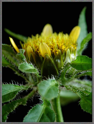
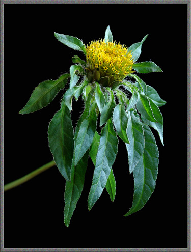
Nodding Beggar-Ticks - Bidens cernuus
The image below shows how different this species appears, with its
long, narrow leaves and attractive yellow petals. When mature,
the flowers tend to droop slightly, as in the photograph, and this is
why the common name was chosen to be “nodding”.
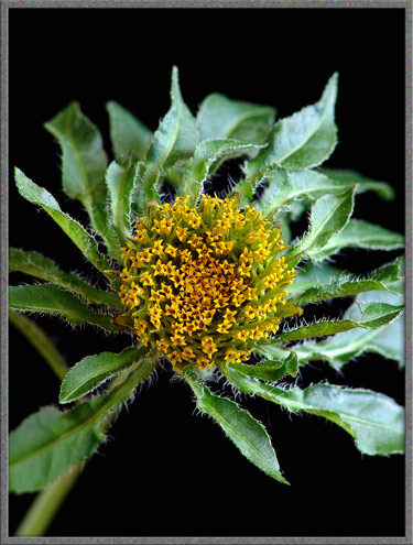
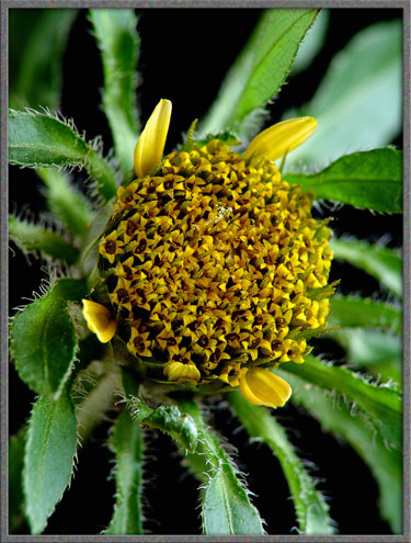
A closer view reveals the distinguishing characteristics of the
species, six to eight yellow ray florets surrounding a yellowish-brown
disk. The outer phyllaries are similar to those of Devil’s
Beggar-Ticks’.
A higher magnification shows that the inner phyllaries, just inside the
petals, are also pale green with dark stripes.
In fact, in this species, the striped phyllary rings extend right to
the centre of the flower-head, and in an immature bloom, form the
central dark area. (The stripes can be seen clearly in the
photograph.) Surrounding this central area, yellow stigmas can be
seen projecting from the flowers.
A side view shows additional details, including a couple of
spider’s-web threads that are commonly found on the flowers.
Two photomicrographs of an immature flower are shown below. At
this stage the tube of fused stamens is visible, each topped by an
anther, but the style has not yet grown out of the end of the tube.
The
final image shows that the pollen grains of this species look
identical to those of the “devil’s” flower.
The identification of the various Beggar-Ticks species was extremely
difficult. There seems to be little information about the plant
available on the Web or in reference texts. My identifications
were based upon the information in the first of the references listed
below. (The others seem to have little or no mention of these
species!)
Photographic Equipment
The photographs in the article were taken with an eight megapixel Sony
CyberShot DSC-F 828 equipped with achromatic close-up lenses (Nikon 5T,
6T, Sony VCL-M3358, and shorter focal length achromat) used singly or
in combination. The lenses screw into the 58 mm filter threads of the
camera lens. (These produce a magnification of from 0.5X to 10X
for a 4x6 inch image.) Still higher magnifications were obtained
by using a macro coupler (which has two male threads) to attach a
reversed 50 mm focal length f 1.4 Olympus SLR lens to the F 828.
(The magnification here is about 14X for a 4x6 inch image.) The
photomicrographs were taken with a Leitz SM-Pol microscope (using a
dark ground condenser), and the Coolpix 4500.
References
The following references have been
found to be valuable in the identification of wildflowers, and they are
also a good source of information about them.
Published in the August
2005 edition of Micscape.
Please report any Web problems or
offer general comments to the Micscape
Editor.
Micscape is the on-line monthly magazine
of the Microscopy UK web
site at Microscopy-UK
© Onview.net Ltd, Microscopy-UK, and all contributors 1995 onwards. All rights reserved. Main site is at www.microscopy-uk.org.uk with full mirror at www.microscopy-uk.net .