A
tour around the 'Micro-Microscope' Model MM02
A field / compact model
with medium to high magnification
originally designed for Indian students.
by David Walker, UK
The Micro-Microscope Model MM02 made by Micro
Instruments (Howrah, West Bengal) was recently drawn to Micscape's attention (see
Acknowledgements). The original design brief was intriguing; see the
two The Times of India
article links below for the background to its design and inventor's
details. A cheap locally made usable microscope was required for Indian
students who may not have access to expensive compound microscopes.
The microscope is being made more widely available as a compact / field
microscope. As standard it offers 100x and 450x magnification with
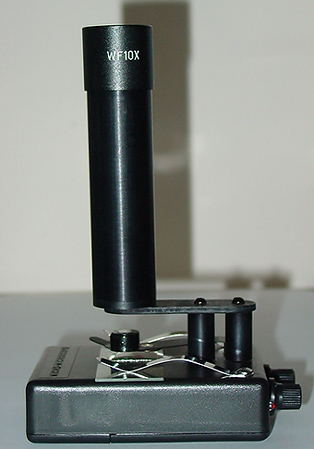
built in variable LED lighting and standard WF 10x eyepiece. Below is a 'tour' of the
microscope and how it performs.
|
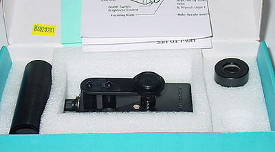
The
outfit includes eyepiece tube, microscope base, eyepiece (10x WF
standard option), instructions and warranty card. (Also a 9V battery if
locally purchased).
When
dismantled the microscope is very compact and could readily be put in
the pocket in a plastic bag to keep dust and dirt off for field use.
With battery it weighs less than 250g.
|
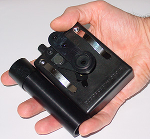
A sturdy plastic box forms
the base. The metal eyepiece tube takes standard size eyepieces which
screws onto a rigid metal plate supported on two metal pillars.
Two spring clips securely hold microscope slides.
Base size ca. 9 x 7 x 2.5cm. Total height 15.7 cm
The rotary knob nearest
camera is a switch and intensity control for the lamp. A
red LED shows when it's on. The rear knob is the focus.
|
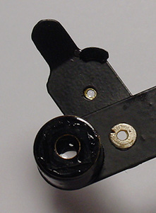
Optics
The two objectives are an interesting design
and described on the maker's box; 'Completely new and very special
objectives have been used. Patent (Pending) technology'. They are
mounted on a stiff springy plate 'P' which is securely fixed at mount
'M'. A black stub 'S' extends down a hole into the base which is the
focus mechanism. The optics swing in a small arc relative to the
eyepiece, but the author couldn't see any deleterious affects in
viewing and focussing. See next section for how the focus works.
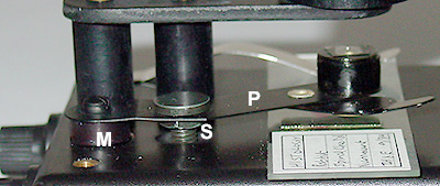
Detail of focus plate and objective support.
A spring stage clip is removed for clarity.
The 10x objective is housed in the small black
casing which has a thin glass plate as dust cover. The 45x
mag is achieved by swinging in the small element, a black stop
aligns it optically. The bottom element by inspection appears to be a
single element plano-convex lens. The side view shows the typical
working distance of the focussed 10x objective. This decreases when the
45x objective is swung in and focussed.
With the standard 10x WF eyepiece supplied
useful mags of 100x and 450x are available. A 15x is available but
don't think a higher mag eyepiece is justified. A 5x eyepiece option
offered would give 50x and 225x which may be better for suitable
subjects and for younger students as less demanding slide manipulation.
The 10x gives a field of view of ca. 1.1 mm and
the 45x ca. 0.22 mm. Test slides aren't really appropriate for a
microscope with its original design brief. In practical
use with real subjects such
as insect parts
and plant / histology sections, the images are competent to the edges,
contrasty and look flat. The 10x does show increasing chromatic
aberration towards field edge. The lamp is plenty bright enough for the
450x.
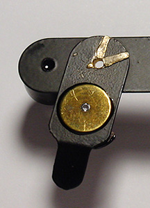
Detail of 10x objective and
auxiliary optic for 45x from above and below.
Right, side view of focussed 10x.
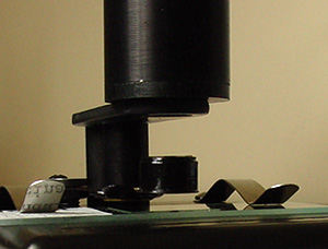
Inside the box - focus and
lighting
Underside with internal case exposed.
The battery compartment is accessible via two
screws and takes a standard PP9 (UK code) 9V battery. For this 'tour'
the glued lefthand compartment was exposed to show the
construction but shouldn't need acccessing as LED's usually have a
very long life. Note that the screw holes are brass bushed to give long
term secure fittings.
The LED seen centre is firmly held in
a metal bush immediately below a small aperture in the plastic box
top i.e. there's no condenser. A potentiometer with dropping resistors
supplies power to the LED.
The focus is a very simple but effective
design. The focus knob turns a fine pitched brass screw whose end
acts on a vertical black rod (actually a screw) with a half
flat to accept it. This neatly translates horizontal
travel to the vertical focus because the black rod is
attached to the spring clip on which the lenses are mounted (see above).
In use
The component parts are simply
engineered for ease of manufacture and to keep cost down but work
together to give a cleverly designed and pleasing little scope. It can
be used in the hand or on a bench and feels stable. I had a lot of fun
with it and would imagine students without normal access to microscopes
would be thrilled with this tool for exploring the microscopic world.
Coupled with the even bright white light that the LED offers, as the
typical images below show, the optics are of sufficient quality
to show what is needed for most biology courses e.g. in botany and
histology. As well as revealing many aspects of live organisms in
temporary mounts e.g. in pond water. I particularly liked the
standard slide layout so slide manipulation is intuitive
e.g. for students, compared with the more awkward inverted
slide requirement for some field microscopes.
The focus works well and feels
tight. The swing-in element is probably best left in when not in use to
keep clean. A simple optics cleaning method is given on the
maker's website. The website also gives one suggestion for
photomicrography with a 35 mm SLR body but the standard eyepieces
should allow other possibilities for imaging e.g. with a suitable low
cost digicam (or mobile phone camera?) that could fit or be held
against the eyepiece.
I believe that the Micro
Microscope is being offered as a portable / field microscope for a
wider market. The reviewer isn't familiar with all available
alternatives and pricing; the only model in production that I'm aware
of that can offer similar mags in the field is the very expensive Swift
FM-31. For potential users requiring such mags in the field at lower
cost and not requiring the optical quality of advanced field or
compound microscopes, the Micro-Microscope could be of interest. The retail
price is $75 (source NG Global).
The images below were
taken with a Moticam 1000 with its relay lens above the supplied
eyepiece.
See links below for examples using a 35mm SLR on the Micro Instruments
website.
|
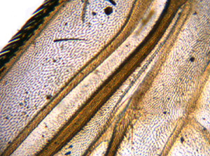
Fly wing detail, prepared slide.
LOMO 7x eyepiece, 10x objective.
|
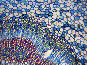
Mexican orange, T/S stained leaf, Biosil slide.
Baker 5x eyepiece, 10x objective.
|
|
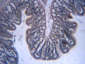
Rat colon, iron haematoxylin and eosin stain,
NBS slide. 10x eyepiece, 10x objective.
|
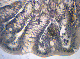
Rat colon, iron haematoxylin and eosin stain,
NBS slide. 10x eyepiece, 45x objective.
|
|
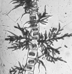
Draparnaldia algae, preserved fluid mount, NBS slide.
10x eyepiece, 10x objective.
|
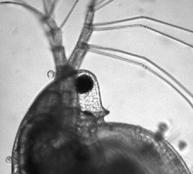
Live daphnia, Baker 5x eyepiece, 10x objective.
|
|
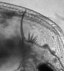
Live daphnia, tail detail, Baker 5x eyepiece, 10x objective.
|
The
waterflea was mounted in a 0.4mm ring cell slide; there is enough room
to focus and move such slides with the 10x objective thus pond
life life studies of some larger invertebrates are doable.
|
Comments to the author David Walker are welcomed.
Acknowledgement:
The author would like to thank Bikash Ghosh of NG Global, Texas, USA
for drawing this interesting microscope to our attention and for
sending a review example which will be duly donated to a good cause
locally.
Links:
'The Times of India' articles. (Note that articles may not be
referring to this exact model.)
'Tiny
microscope at microscopic price'.
June 2002.
'CM
bowled over by mini-microscope, praises inventor'. September 2003.
Maker's Micro Instruments website links:
Please note: There is javascript on the maker's 'Home' page which
gives a (hopefully) false positive with some virus checking software
and which may prevent access. The web site owner assures me it is
benign script but visitors may wish to avoid clicking the 'Home'
button. Other pages on the website don't have this script and direct
links are given below.
Product
details.
Example
images from 35 mm SLR camera.
Maker's
and stockist contact details.
© Microscopy UK or their contributors.
Published in the August 2005 edition of
Micscape.
Please report any Web
problems or offer general comments to the Micscape Editor .
Micscape is the on-line monthly magazine of
the Microscopy UK web site at Microscopy-UK
© Onview.net Ltd, Microscopy-UK, and all
contributors 1995 onwards. All rights reserved.
Main site is at www.microscopy-uk.org.uk with full
mirror at www.microscopy-uk.net .