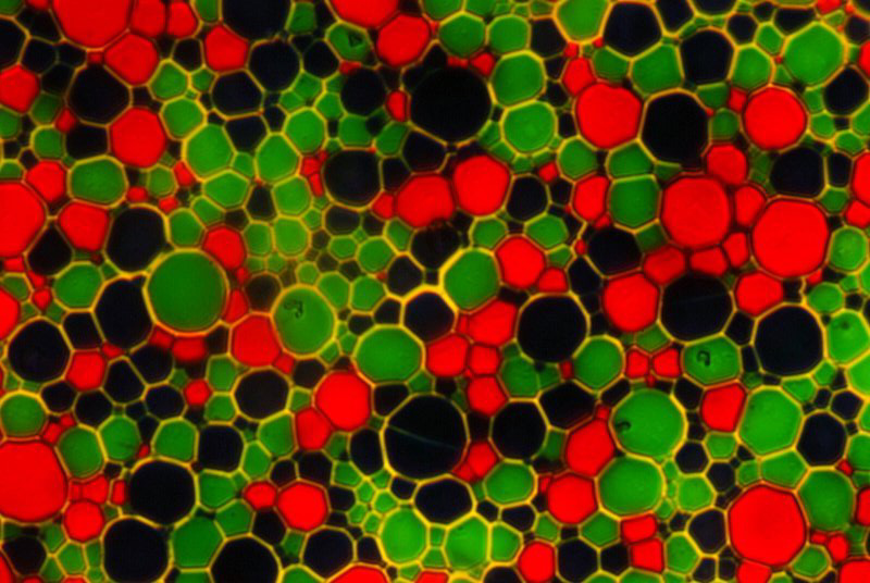
Fig 1 Agfa photographic process very early 20 century. The first photo taken with the D200 camera.
|
A Tale of Two Cameras Dan McNeil USA |
For about the last decade I have used a video camera and a Snappy interface to make digital images with my microscopes. I would then import them into word and annotate them before printing on high quality photo paper. The photos looked nice when printed at 4 inches by 6 inches but started to fall apart when printed larger. This was rather frustrating for someone who liked to make large prints from 4” X 5” transparencies and negatives so I started to investigate dedicated digital cameras for microscopy. Most cameras of this ilk were outrageously expensive or merely modified video cameras similar to what I had been using. After much browsing of the internet I narrowed my search to two cameras which made huge files and hence large prints by moving the sensor in small increments during exposure. One of these was American made, only moderately expensive, and had received some favorable publicity on the microscopy website (USA) so I ordered one. For reasons which will soon become apparent I will refer to this camera as Brand X .
Brand X arrived and I quickly loaded the software and mounted the camera on my microscope. The first object I attempted to photograph was a slide of an early 20th century color photographic process consisting of red, green, and blue spheres. The image appeared on the screen as uniform dirty yellow. I tried fiddling with the color sliders on the control page but after several hours was unable to achieve a satisfactory coloring of my image. I was also unable to get a really sharp image. This problem carried through regardless of the type of specimen. Diatoms looked especially gruesome. All in all the images obtained with this camera were inferior to those obtained with my video/Snappy setup. After some email correspondence with a sales rep he came to my house and fiddled with my setup. His results were certainly better than mine but still not something that I would like to present as my work. Colors were off and sharpness left something to be desired. The necessity to diddle with color bars and sharpness issues seems to go against the Microscopy Society of Americas guidelines for ethical presentation of digital images (ref. 1). Brand X was a big disappointment and was soon put aside in favor of the Snappy/video setup.
Recently I purchased a Nikon D200 single lens reflex digital camera. This camera has a removable lens and a 10 mp sensor. I thought it might be a good microscope camera as well as a general purpose tool. The very first photo I took with it was on my microscope of the red, green, blue spheres. I used the settings that were on the camera as received and made no changes at all. To my surprise color was right on the money with acceptable sharpness. My first photo with the Nikon is presented in fig. 1. I quickly changed the specimen to a diatom slide and was again pleasantly surprised with the results. Again no changes to the camera settings were made. Figs. 2, 3 and 4 are of the diatom Coscinodiscus made with the Nikon D200 camera.
Fig
1 Agfa photographic process very early 20 century. The first photo
taken with the D200 camera.
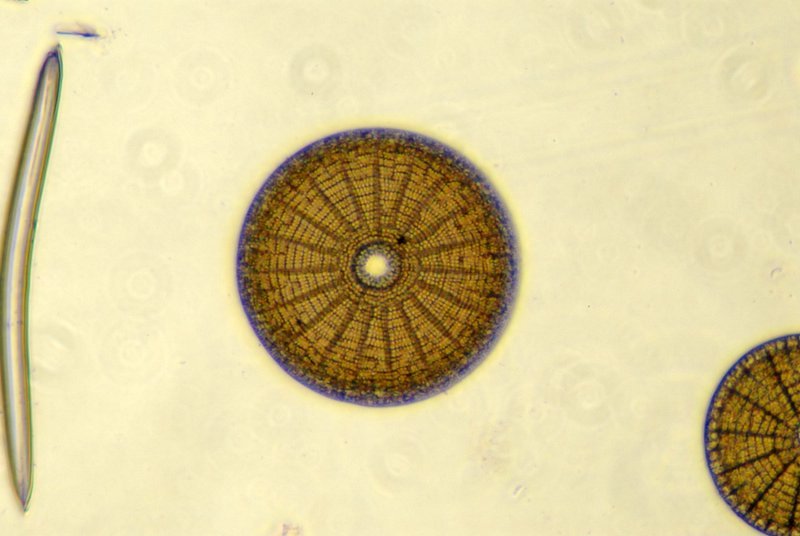
Fig
2 Diatom illuminated with achromatic condenser and photographed with 32X
objective.
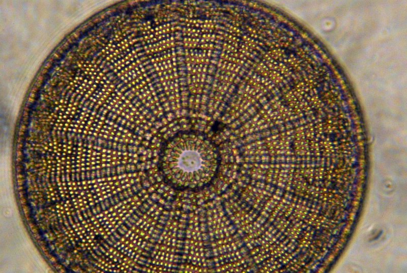
Fig.
3 Diatom illuminated with Heine condenser and photographed with 50X objective.
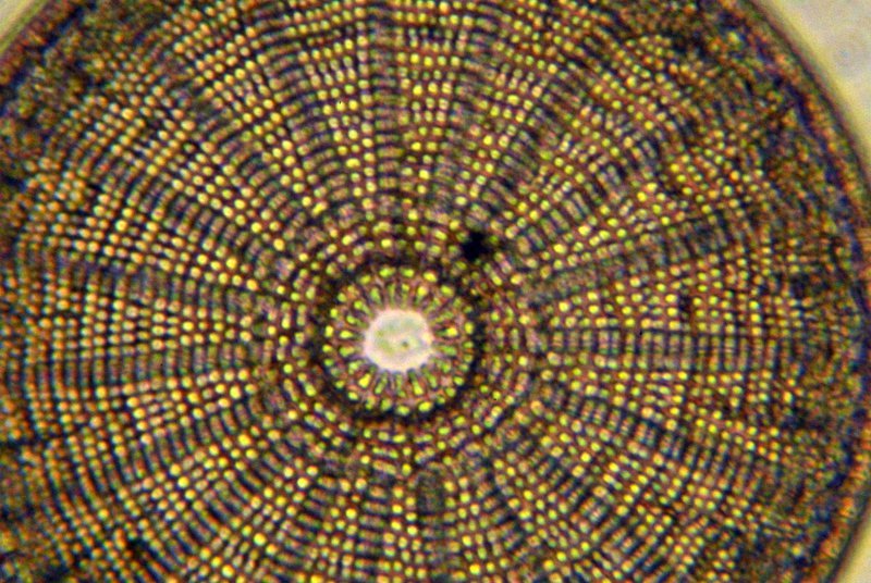
Fig.
4 Diatom illuminated with Heine condenser and photographed with 63X objective.
Adapting the D200 to my microscope was easy since I used the same setup proven for ten years with my Nikon F3. This consists of a small support stand, a machined aluminum tube attached to a T ring and a fabric light lock. The tube fits very loosely over the phototube of the microscope and has some lateral movement for alignment. It is provided with coarse threads along its entire inside length to act as baffles against stray light. It is coated inside with Krylon ultra flat black paint. For a light lock I use a two layered lint free black fabric tube with drawstrings at each end made for me by my daughter. The support stand isolates the camera mirror vibration from the microscope and maintains alignment. For really critical shots I use the mirror lock up feature and the self timer option to completely eliminate camera shake. By raising or lowering the height of the camera I can make it almost parfocal with the visual microscope. My setup is shown in fig. 5. [The camera shown is my F3 which is only slightly different in appearance from the D200 which is being used to make this picture.] To import the photos into the computer I remove the memory chip and insert it into the computer. This is much faster than downloading from camera via USB.
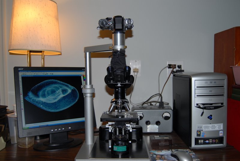
Fig.
5 The author’s photomicrographic setup for film. The D200 camera was used
to make this photo.
One problem I have with the D200 camera is focusing through the viewfinder. Unlike the F3 which has interchangeable screens the focusing screen and Fresnel lens interact with the apparent “point” source of light from the eyepiece to create moire making it difficult to see very small features. I get around this by taking several different images at different focal planes and then selecting the best image when I view them on my computer screen. I then erase the not-so-good images and use the space for something else. Fortunately digital storage space is cheap and reusable. I am currently investigating using the cameras LCD monitor for critical focusing.
The largest file size can be printed 13” X 19” at 200 dpi. While still not the equivalent of film, digital technology has the advantage of showing virtually instant results and can produce only one photo without wasting the remainder of the roll (and money) as I have often had to do in the past with film. It is also constantly getting better.
Any questions or comments can be directed to the author Dan McNeil.
Reference
1) J. M. Mackenzie, M. G. Burke, T. Carvalho and A. Eades; Ethics and Digital Imaging; Microscopy Today; January 2006; pp 40-41.
Microscopy
UK Front Page
Micscape
Magazine
Article
Library
Published in the August 2006 edition of Micscape Magazine.
Please report any Web problems or offer general comments to the Micscape Editor.
Micscape is the on-line monthly magazine of the Microscopy UK website at Microscopy-UK
©
Onview.net Ltd, Microscopy-UK, and all contributors 1995 onwards. All
rights
reserved. Main site is at www.microscopy-uk.org.uk with full mirror at www.microscopy-uk.net.