|
|
A
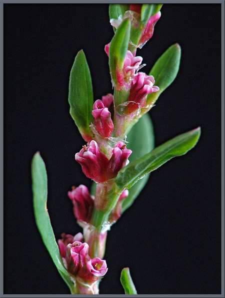
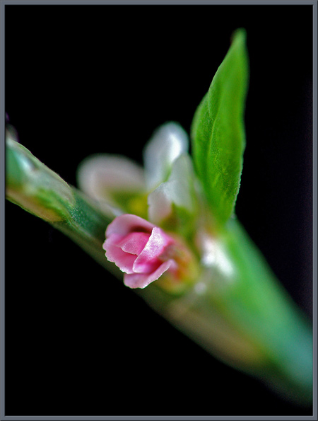
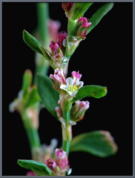
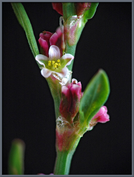
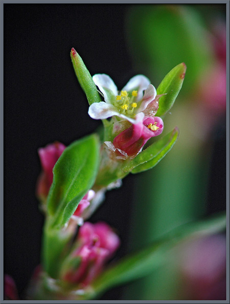
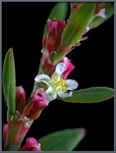
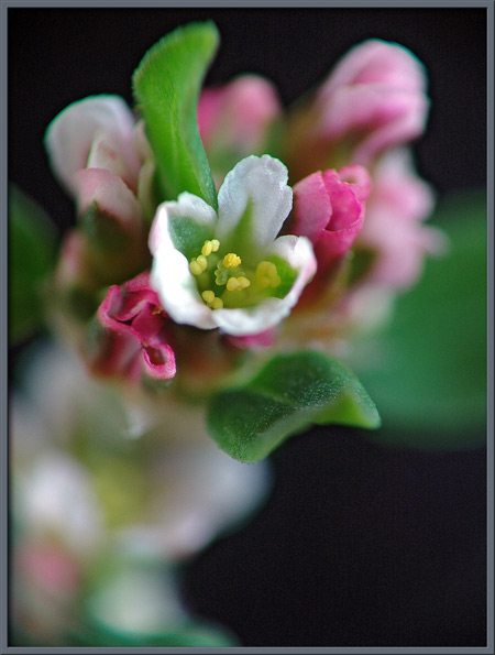
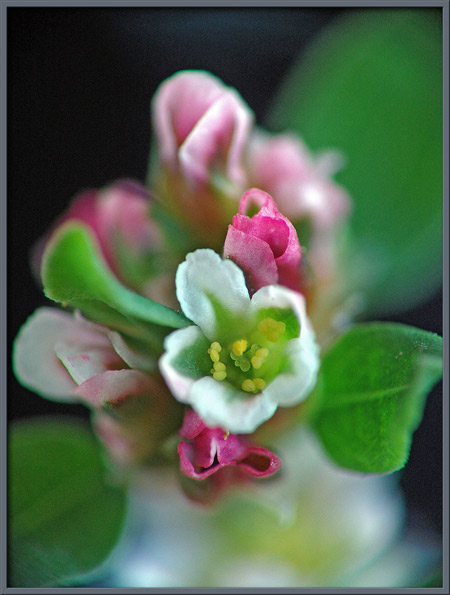
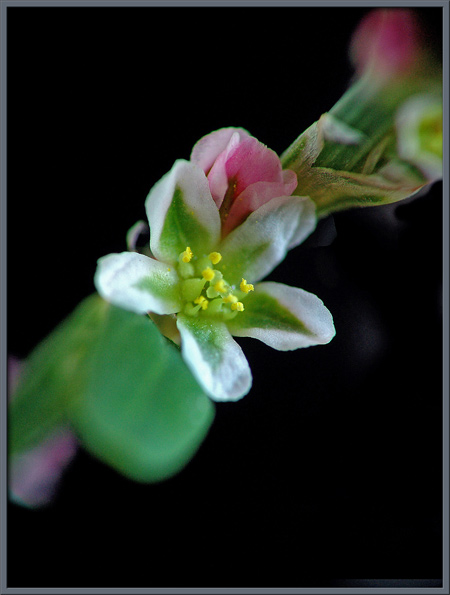
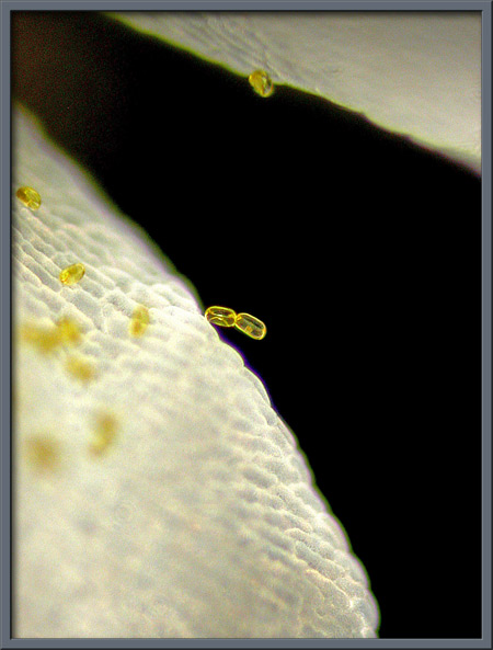
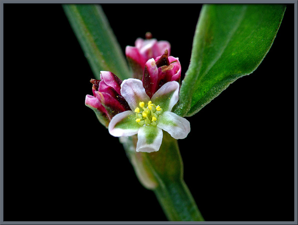
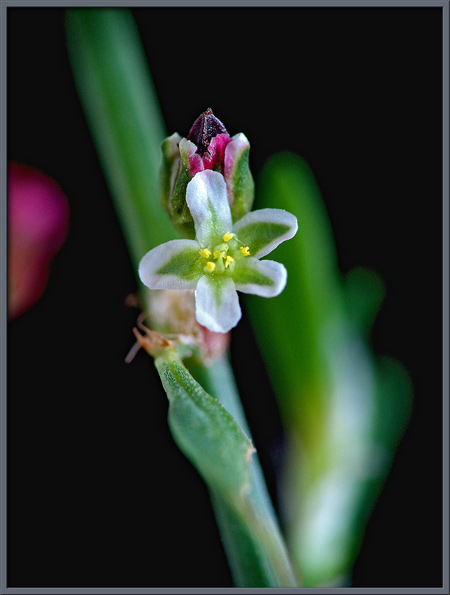
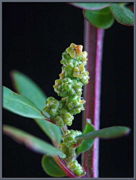
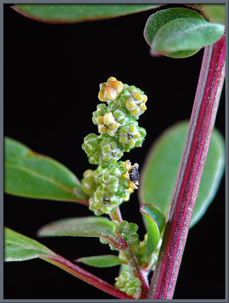
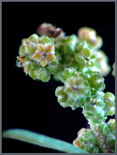
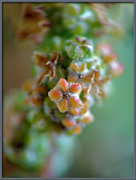
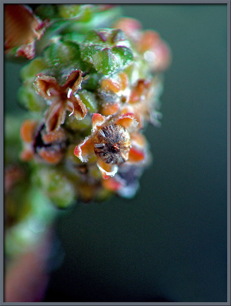
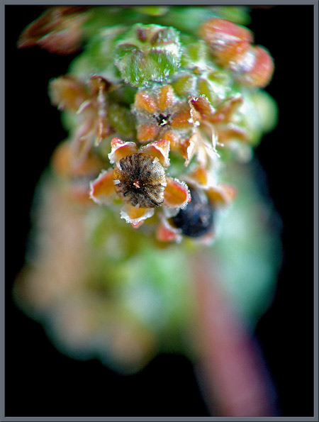
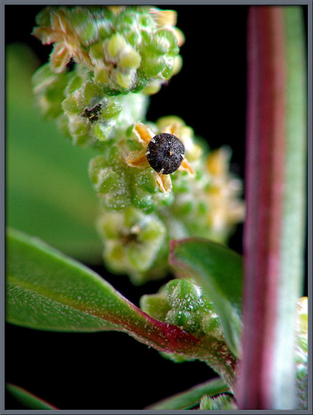
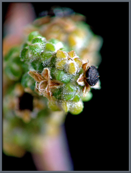
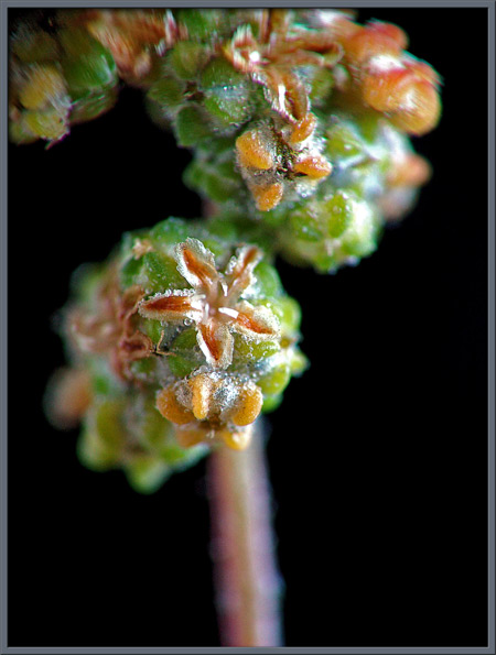
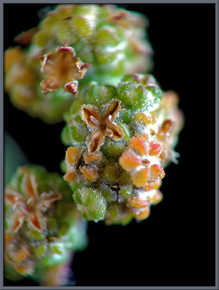
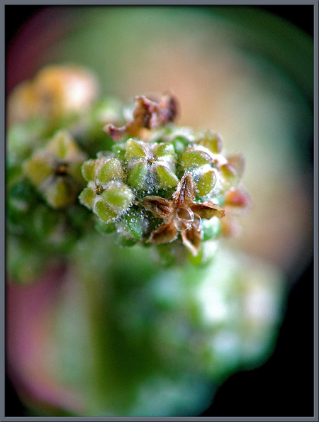
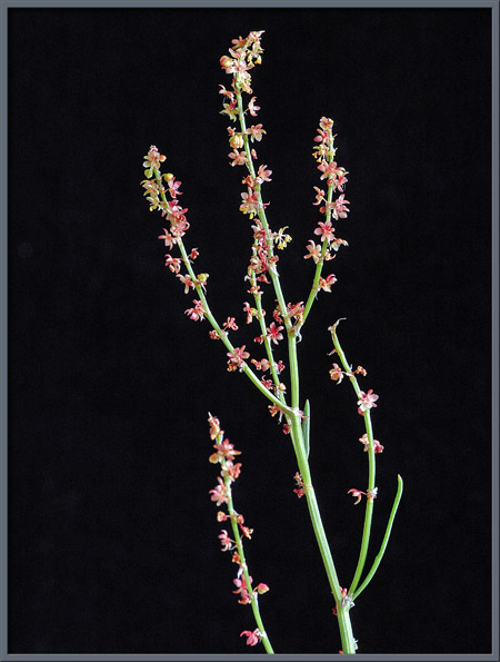
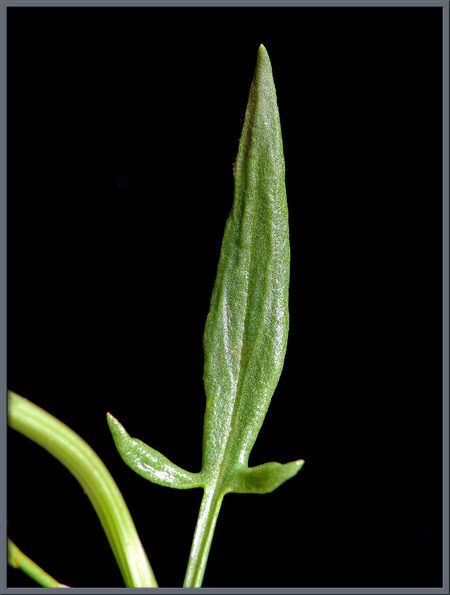
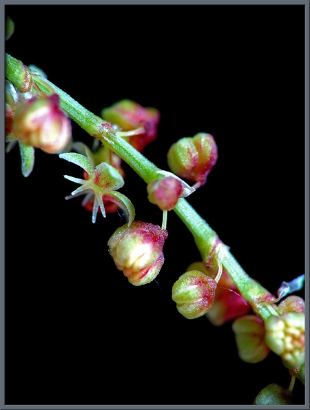
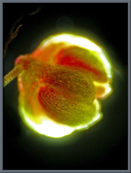
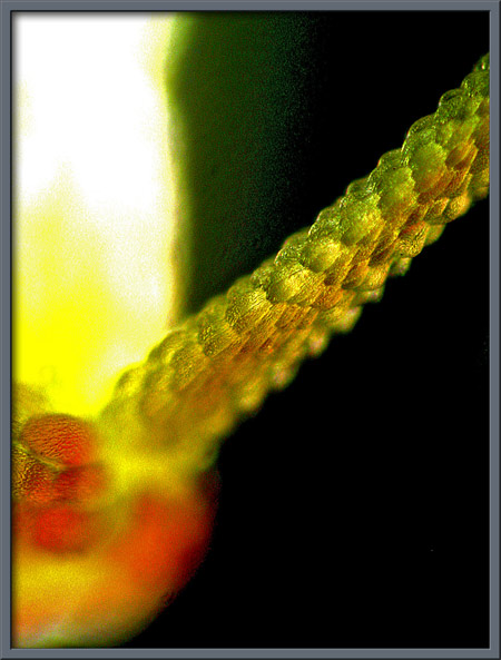
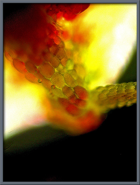
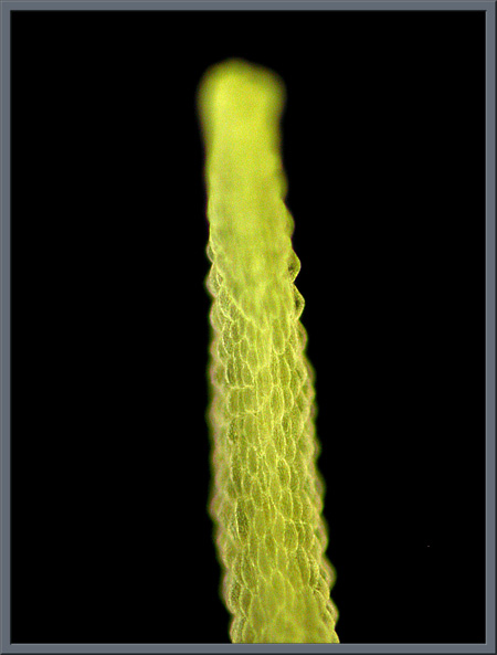
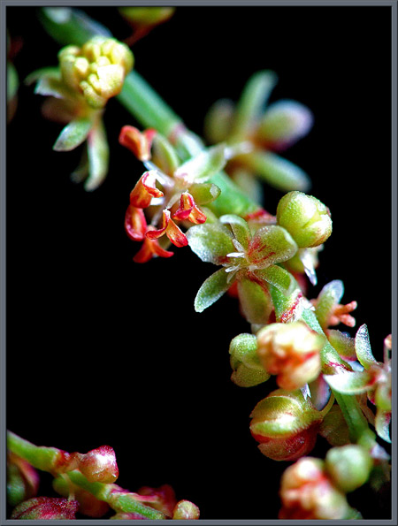
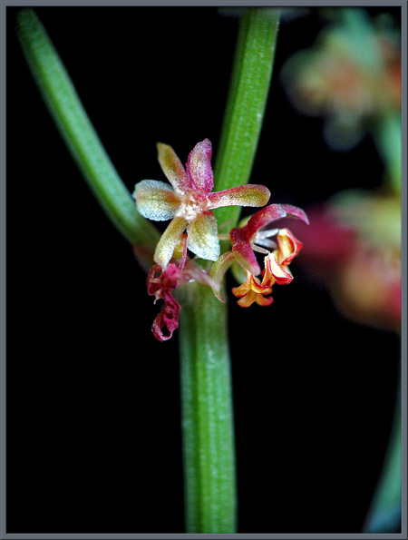
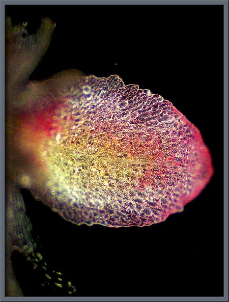
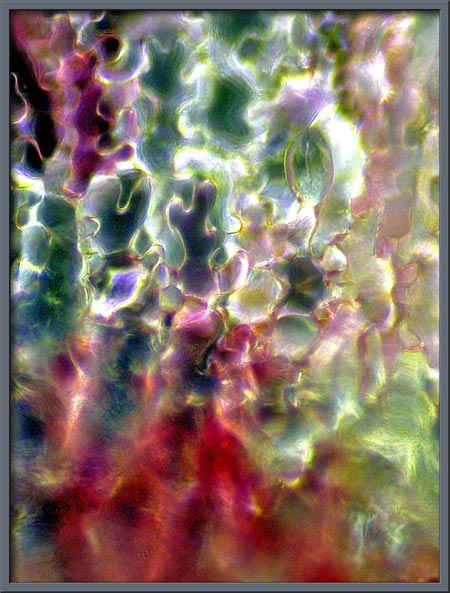
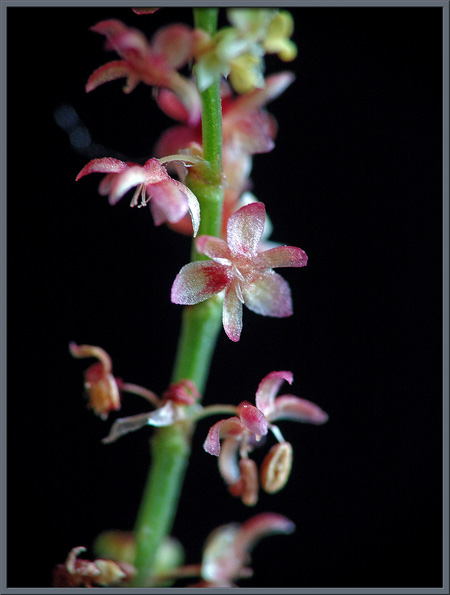
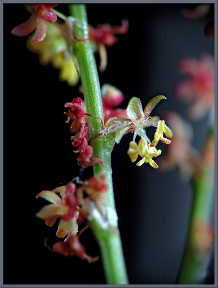
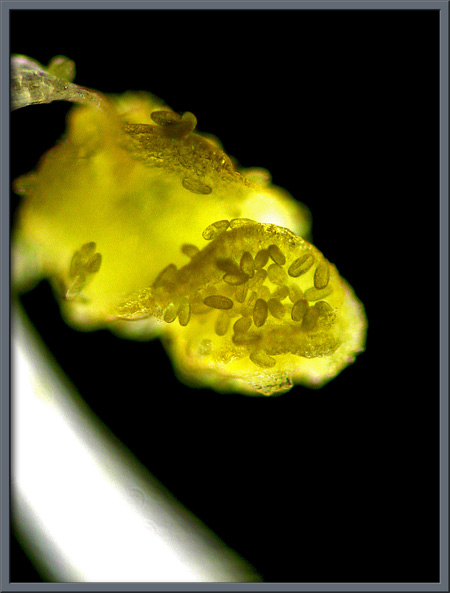
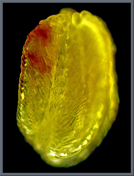
Close-up View of Three Extraordinarily Small Wildflowers
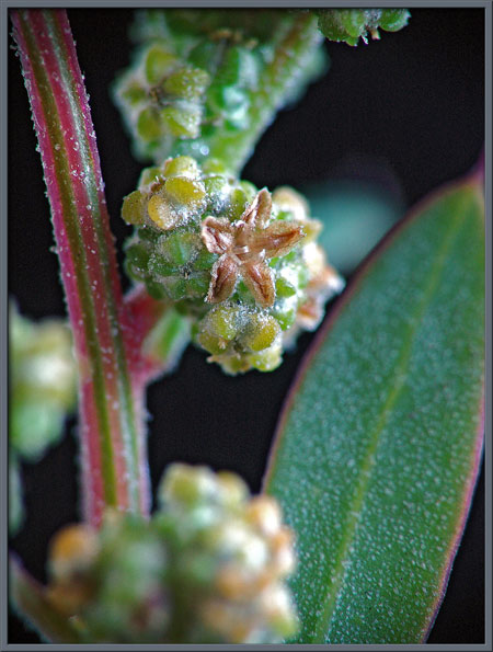
Common Lamb's-Quarters (Chenopodium album) Sheep
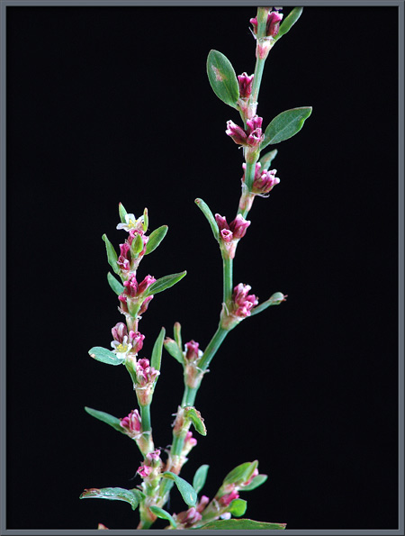
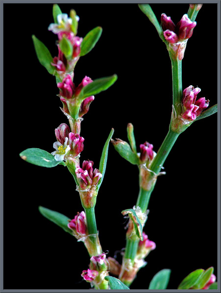
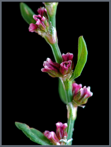
Sorrel (Rumex acetosella)
by
Brian Johnston (Canada) |
|
|
A
Close-up View of Three Extraordinarily Small Wildflowers
Common Lamb's-Quarters (Chenopodium album) Sheep
Sorrel (Rumex acetosella)
by
Brian Johnston (Canada) |


Published in the August
2007 edition of Micscape.
Please report any Web problems or
offer general comments to the Micscape
Editor.
Micscape is the on-line monthly magazine
of the Microscopy UK web
site at Microscopy-UK
© Onview.net Ltd, Microscopy-UK, and all contributors 1995 onwards. All rights reserved. Main site is at www.microscopy-uk.org.uk with full mirror at www.microscopy-uk.net .