Coins and Microscopes
Robert Pavlis, Girard, Kansas USA
Coins can make very interesting subjects for microscopes. Numismatists are always very concerned about the condition of coins in their collection, and microscopes make it possible to see all flaws in great detail. Microscopes also permit examination of surface characteristics of the metal employed in making the coin. Here we will examine two very different coins, the first is an interesting Icelandic coin that commemorates the 1000th anniversary of the founding of the Icelandic parliament. The second is an ancient coin, Sear-173, minted between 119 and 110 b.c. that shows the god Mars on the obverse, and horses on the reverse.
Until the 20th century people tended to regard coins as being certified pieces of metal that were guaranteed to contain a standard quantity of gold or silver at a standard fineness. They tended to regard base metal "coins" only as tokens, indeed in many countries base metal "coins" were not even legal tender and could only be used for making very small purchases. The development of fiat (worthless) money has changed things dramatically so that "coins" in the old sense of the term no longer circulate. Many governments, however, still make gold and silver coins that are available for those who want to avoid having their fiat money inflate into oblivion. (The people who purchase these do have a point. Inflation in the US in the last 100 years has reduced the value of the dollar so much that a modern dollar buys approximately the same as a cent then. In the UK the value of the pound has also fallen, so that 1d 100 years ago is roughly equivalent to a modern pound! This makes the modern pound about 1/240 the value of one 100 years ago.)
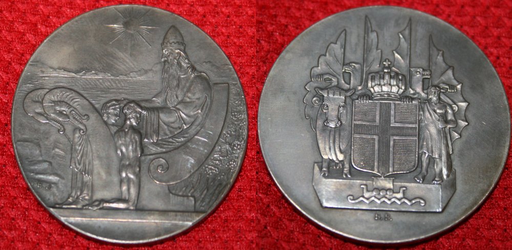
1930 10 Kronor Icelandic Parliament Commemorative
Many years ago I attended an auction where I obtained an interesting Icelandic coin that commemorates the 1000th anniversary of the founding of the Alþing or Althing, the Icelandic parliament. Many numismatists contend that this is not a coin because the denomination, 10 kronor, is only printed on the edge of the coin. This coin does not appear in many coin catalogues because of this fact. It is sometimes referenced as KM-M3.
What is amazing about this coin is the fact that it has no legends on either the reverse or obverse--only the denomination on the edge. Indeed, the only letters are some very small letters that appear to be designer's initials. Although the coin was issued in 1930 there is no date on the coin, and although it commemorates the founding of the Alþing, there is no legend to indicate that fact! In fact, nothing really connects it with Iceland!
The two images immediately below this text show the obverse and reverse sides of this remarkable coin:
Since the coin is quite large, its diameter is 45 millimetres, the images above were obtained using a Canon digital single lens reflex camera with its standard lens.
The remaining images were taken with one of two systems. The first system utilised a LOMO MBS-10 stereo microscope equipped with the standard LOMO photo attachment and the same Canon digital SLR used above. (This requires the use of a M42 to Canon adaptor.)
The second system utilised the same Canon digital SLR as the camera and an Olympus BHM microscope equipped with trinocular head and an Olympus 2X photo ocular. Only metallurgical objectives were used for the images shown here.
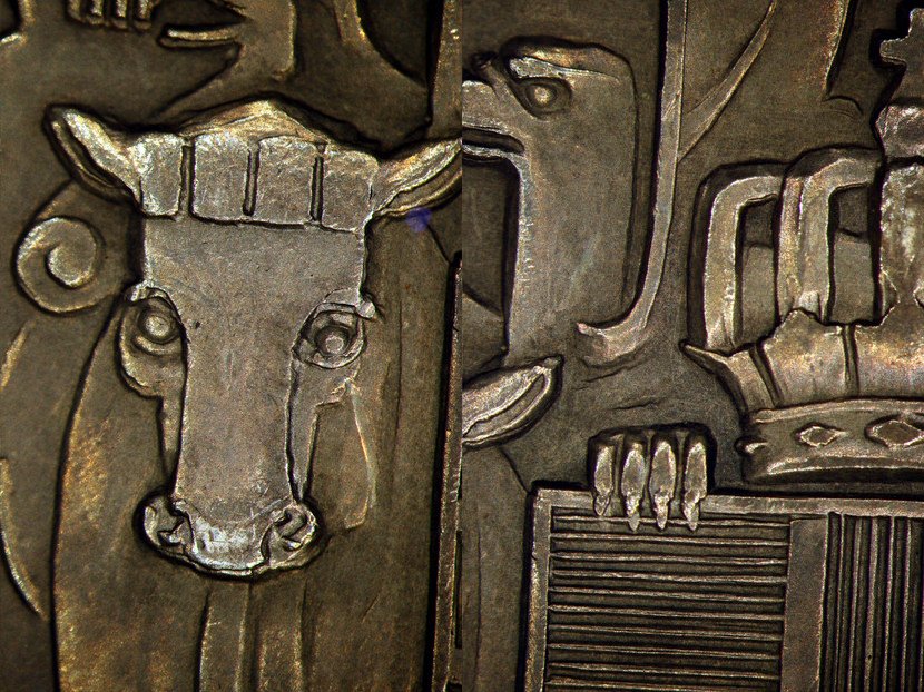
The image below and to the left is of the Ox that can be seen in the image of the reverse side of the coin in the image above. The image to the right shows the ox's horn, along with the small birds and the base of the large crown. Part of the large bird in the background is also visible. These were both taken using the LOMO MBS-10 and its 2X objective.
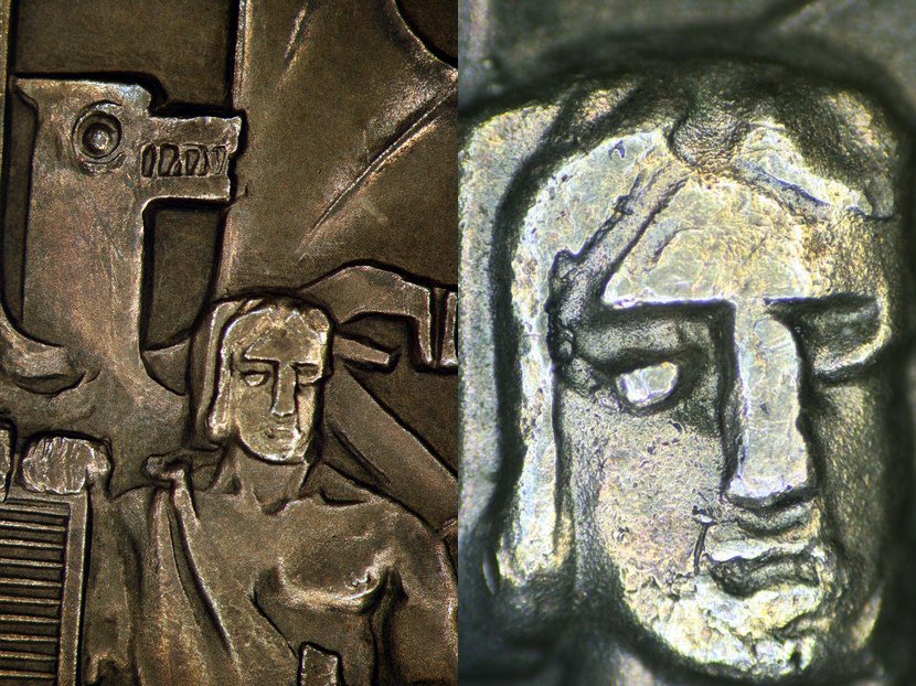
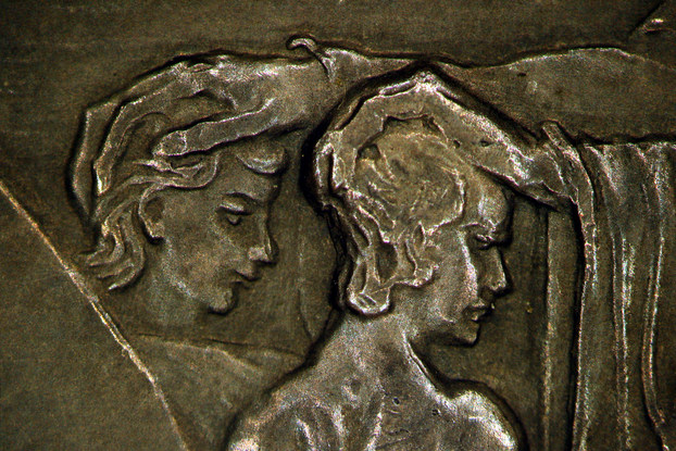
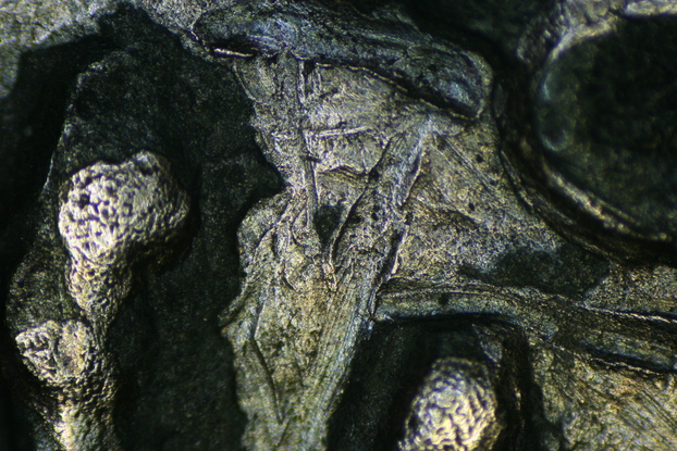
The image below shows the dragon like figure and the warrior on the left side of the coin's reverse using the LOMO MBS-10 and 2X objective. The image at right was obtained using the Olympus BHM with a 4X objective with "dark field" illumination. Note how the metal is beginning to appear very granular at this power.
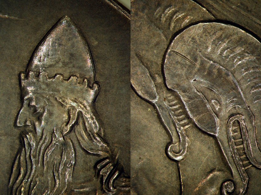
The image below shows the King of Thule and the dragon like figures on the left side of the obverse of the coin. They were also taken with the LOMO MBS-10 and 2X objective.
The next image shows the kneeling subjects. When one examines this coin using a stereo microscope it is apparent that this coin has never been circulated, and has never been stored under conditions where it could have been allowed to rub against other coins. There are practically no scratches visible at all.
The next image was taken of a flat area of the coin utilising the Olympus BHM microscope with the same 2X photo ocular but with a 40X objective. This image was obtained with the metallurgical equivalent of dark field illumination. Note the granular structure of the metal that is visible at this magnification. This coin is remarkable in its lack of scratches, one can scan the coin at this power and find only a few very minor scratches.
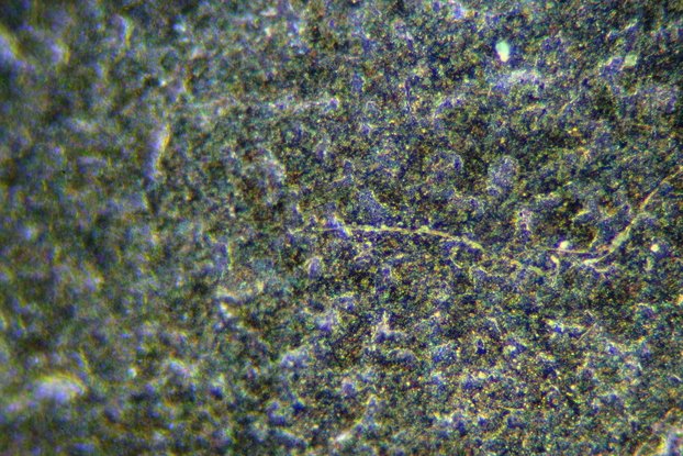
The image below is of a similar area as the above image, except it is obtained with the metallurgical equivalent of bright fieldwith illumination through the objective. Note how the grain structure of the metal is very prominent here.

All the images shown above are of the same coin. Microscopic examination of coins can certainly reveal many things about them that are not visible to the unaided eye. The designs and condition of the metal tends to vary quite dramatically from one coin to another. The next section shows a rather dramatically different silver coinan ancient Roman denarius.
Roman Republic Mars and Horsemen Denarius, S173.
The images below are taken with the LOMO MBS-10 using the same system described above. The first images show the full coin using the 0.6X objective. This objective exhibits substantial curvature of field, but the images are still quite good.
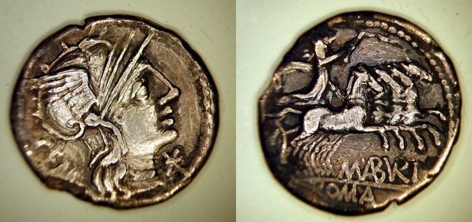
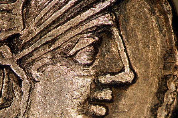
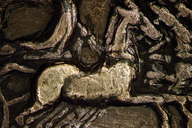
This silver denarius coin is about 2125 years old. Its diameter is approximately 20 millimetres. The image below was obtained using the same LOMO MBS-10 setup, but with the 2X objective. It shows a closeup view of the face of the Roman god, Mars.
The next image was obtained using the same conditions that were used for the above image, except that the coin was flipped over to show a close-up of the horses.
The atoms that form the metal structure are in constant motion. Over periods of hundreds of years the crystalline grain boundaries that form as the metal atoms diffuse from one spot to another cause long term physical changes in the coin. The exact consequences of this depend mostly on original chemical composition. Because ancient Roman coins differed somewhat in composition, the consequences of this vary from coin to coin.
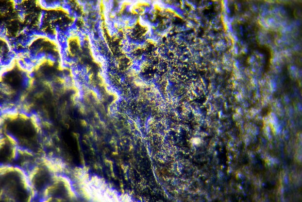
The image below was obtained using the Olympus BHM with the 4X metallurgical objective. It was obtained using the metallurgical equivalent of dark field. Notice the even at this low power the surface of the coin appears quite granular.
The image below was obtained using the Olympus BHM with the 40X metallurgical objective. It was again taken using the metallurgical microscope equivalent of dark field.
The next image shows a field in the same area of the coin with the metallurgical equivalent of bright field.
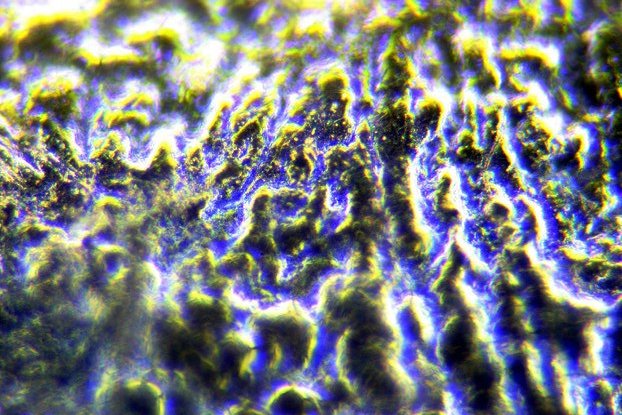
The appearance of the silver is dramatically different in the modern Icelandic coin and the ancient Roman one! If the ancient Romans had had microscopes and had obtained images of the same area of the coin the appearance would be dramatically different than it is today. The segregation of grain boundaries actually makes old coins such as this brittle so that they can shatter when subjected to shock!
All comments to the authors via Robert Pavlis are welcomed.
Microscopy UK Front Page
Micscape Magazine
Article Library
Please report any Web problems or offer general comments to the Micscape Editor .
Micscape is the on-line monthly magazine of the Microscopy UK website at Microscopy-UK