|
|
| From Brightfield To 'Smoke Rings'(Part 3)
A Personal Odyssey By Paul James. |
Continuing the saga of convoluted trains of thought concerning illumination ............. Part 3 starts with some consideration of theoretical improvement of imaging through the telescope which eventually led me to discover some enhancement of medium/low power imaging from COL.
I remember at one time wondering what the effects of fitting an annulus over the aperture of a telescope might have on the image. I reasoned that since wider telescope objectives/mirrors resolve finer details than narrower examples, the actual high imaging potential must stem from the outer zones of the objective/mirror, since all telescopes share the properties of a narrow inner zone. So I made a simple cardboard annulus of about 140 mm diameter with about 8 mm radial air gap and placed it over the 150 mm objective of the telescope. The resulting imagery of the moon was expectedly dimmer, but the image was decidedly softer than without the annulus in place. The craters and other rugged features were fairly sharp but this acuity appeared to be artificial, and the smoother areas, the plains, looked muddy and lower in contrast. Clearly it seemed to me that diffraction was to blame because the annulus had 2 diffraction inducing edges each side of a narrowish air gap. Thus any chance of scrutinising the image from selected zones of the telescope's objective was compromised very effectively by diffraction. Since all objective/mirror assemblies must have a hard edge of some sort at their periphery, the diffraction so generated within the normal full aperture image, seems to be an insignificant part of it, or at least the degrading effects are masked or disguised by the rest of the imaged light that the full aperture generates. Star imagery however elicits diffraction's artifacts very effectively since the first couple of concentric rings so produced ( Airey's rings ) can be seen at higher powers unimpeded over the dark sky background.
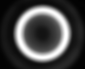
The natural consequence of this simple experiment in practical telescope imaging for me was to wonder if the diffractive component could be nullified in any way, and if it could then the resulting eradication of the Airey's discs that diffraction produces around all points of detail etc., would undoubtably improve imaging contrast. But the designer of optical instruments would probably assert that this wishful pipe dream is unassailable. Nevertheless the idea of having a 'soft edged' annulus in front of the objective/mirror cell came to mind. The illustration below is the sort of annulus I'd imagined, where both sides of the air gap became very gradually opaque, so theoretically defeating the generation of diffraction! It would probably look like the 'smoke ring' illustration below:-
|
|
A large optically flat glass disc having the necessary 'dye' or light absorbing super fine, homogenously integrated colloid, might hopefully suppress diffraction because of the complete absence of a hard edge. Nevertheless it was this rather lofty idea that gelled some parallel thinking in microscopy. In fact I wondered what would happen to COL imagery if the annuli generating it were to have 'softened' edges......ie having a lower capacity for generating diffraction?
However in microscopy the diffraction generated below the specimen by the lens mounts and iris diaphragms in BF does not directly end up spoiling the final image since the specimen itself seriously disrupts the path of diffracted light from the annulus/source. The only really significant diffraction generated by the specimen itself of course occurs around its more opaquely rendered boundaries. Ironically of course without the lower order diffraction that's generated from the internal boundaries of cellular structure and the like, the microscope's brightfield image would be of little use.
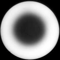
So out of sheer curiosity I conducted some simply trials using small coverslip diffusers in close proximity to the COL annuli in the substage condenser. The idea was to bring about an annulus that looked soft edged through the phase telescope. Deploying such a diffuser of ground glass over the astronomical telescope's objective would naturally result in destroying the image entirely. We could however easily 'shred' the lamphouse's output of light in the compound microscope with such a diffuser, and therefore simulate albeit rather crudely the ideal soft edged annulus. Depending critically on the spacing of the diffuser above the annulus, the appearance of the desired halo of light, the 'smoke ring' I allude to in the article's title, looked interesting :-
|
|
|
|
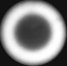
Left image is phase 'scope view of a 'diffused' annulus which includes the outer hard edge masking of the objective . The image right is similar but the na of the ring is a little less so the diffuser's outer soft edge is revealed. |
I have experimented with the diffuse annulus technique now for a while with a number of different objectives, condensers, various annuli, varying grit sizes to frost the glass, as well as altering the placement of the diffusing screens. The eyepiece view was very similar to normal COL, but there was a distinct reduction in unwanted imaging artefacts across the entire field most especially between x10 and x3 objective usage. Eye positioning above the bino head became a little less critical too, and the objectionable inner hairy edge field zone disappeared completely leaving a 'normal' and even field similar to high power COL. Above this level when using x20 objective and higher powers the diffusing screen serves no useful purpose as the field is generally even and very detailed in normal COL anyway.
|
|
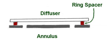
Of course the diffuser needs to be above the annulus to 'soften' it's edges. The space between the annulus and diffuser shown above is the very minimum necessary for effective success, with greater distancing ideally required. This can be a problem because there are physical limitations to where we can site the diffuser. Fliptop condensers have accessible space for a diffusing screen between the top element and the lower body which you might find ideal. Placement on the top lens of the condenser isn't really effective, nor practicable, as is trying to fit one between the lens elements of a normal condenser. The annulus could be lowered instead since in lower power COL its ideal siting at the anterior focal plane of the condenser isn't so critical as with higher powers.
|
|
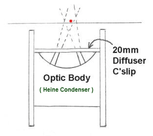
Trials with the Heine condenser revealed the same improvement of the image with medium to low power objectives too. Diffuser placement however was just as critical. I started with a 20 mm coverslip (ground with 400 grit carborundum) which slipped into the Heine's barrel conveniently as illustrated above. In practice the condenser needed to be a long way down below the specimen to be effective. Nevertheless its objectionable field artifacts at extreme low power down to x3 objective in brightfield ( COL ) mode was nullified completely with this simple 20 mm diffuser. More output power from the lamphouse might be an advantage, since the diffuser reduces light intensity which some might find unacceptable.
ODDS AND ENDS
These very brief notes will hopefully give enough of the basic ideas so that those practitioners of COL can follow through with their own experimentation to hone or fine tune these notions for their own particular ends. Be mindful of the grade of grit required for 'frosting' glass for diffusers, ideally leaning toward the finer grades of carborundum 300 - 400 or finer. Coverslip diffusers are quickly ground, but have the tendency to crackup during the process.....ie when rubbed over another glass substrate . Use a well wetted glass base plate for this and use a tiny pinch of grinding powder laced with a drop of soapy detergent. More robust squares of glass salved from broken slides can be used too. Their rough edges can be smoothed at the same time as frosting their surfaces.
Even if you think my theories are tinged with anomalies.... this halo of light.....the smoke ring, actually works well yielding very smoothly represented low power imagery, ideally suited for entomological studies yielding fine contrast without any of those strange artifacts that put a lot of folk off the low power COL/annular lighting concept. The only real problem is finding the right substage location (elevation-wise) for the diffuser where it can do its business, as well as being able to be removed and replaced conveniently. If it looks good through the phase telescope or Optovar then clearly you are close........finally checking through the eyepiece of course.
CONCLUDING THOUGHTS
As amateur microscopists we ought to be mindful of the astronomers' plight. Their imagery generated by the telescope originates from surface illuminated planets and self illuminating stars and nebulae etc.. In short they cannot alter the light that the telescope gathers to image their prey, but can and do modify it after imaging with filters etc.. We microscopists have the great advantage, that of being able to apply different lighting conditions on our prey. Only one particular situation in astronomy occurs where there are significant light changes before reaching the telescope, and that of course is the moon's constantly changing phases. Here the light varies from 'oblique' to 'vertical illumination' which elicits highly contrasting effects that floods the images with richly rendered detail. By comparison the amateur microscopist can have a field day regarding the illumination of a specimen, limited only by his imagination. Brightfield is the bedrock lighting technique with many variants .......... one or more suiting the personal visual appetites of each individual amateur microscopist. The desired image is simply what you can contrive from the specimen and the illuminant.
| All comments welcome by the author Paul James |
Microscopy UK Front Page
Micscape
Magazine
Article
Library
Published in the August 2009 edition of Micscape.
Please report any Web problems or offer general comments to the Micscape Editor.
Micscape is the on-line monthly
magazine of the Microscopy UK web
site at
Microscopy-UK