|
Spring Pond Harvest: A Title Which Has Very Little To Do With The Contents Of This Essay by Richard L. Howey, Wyoming, USA |
Each spring there is a certain turning point when I know that it is time to get out my nets and collecting bottles and head for the nearest pond. After many years, I have learned to sense when that burst of life and energy will appear and a rich variety of forms will be actively producing a harvest for my nets. For me, the ideal times to collect are the spring and autumn or what I call the pre- and post-mosquito seasons. In early and midsummer, they occur in oppressive swarms around open water and, of course, now there is the addition of the potentially serious discomfort of West Nile fever but, in the spring, the explosion of aquatic protists and invertebrates makes every trip into the field an exciting adventure. These waters are filled with marvels and mysteries that one could only begin to address in a hundred lifetimes.
Every year, I set out with a sense of anticipation and joy, for every year I have found organisms that I had never previously seen and I have had the fun of trying to study them and identify them and, if I get lucky, produce some good photographs of them. Part of the joy is the challenge for each time, I am aware that I am entering a world so marvelously alien that it convinces me that all of the really good science fiction writers are or were closet microscopists. What astonishes me over and over is that here is a multitude of hidden universes all around us which 99% of the world’s population is totally or nearly completely unaware of. I like to think of the amateur microscopists and natural historians as explorers who have before them the exhilaration of inexhaustible adventures. The fact that others have seen, described and studied the splendid desmid Micrasterias in no way detracts from my elation in observing and investigating it.
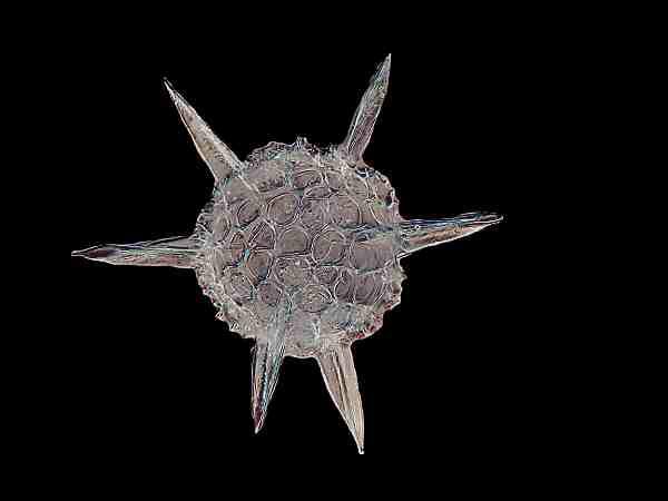
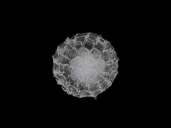
There are many examples of such elaborate kinds of geometric forms in nature and so it is not surprising that historically God was often thought of as a mathematician. Consider the marvelous radial symmetry of radiolaria. Of course you won’t find these in ponds, since they are exclusively marine.
This sort of lovely mathematical precision can be found in heliozoan amoebae such as Actinosphaerium.
Also a similar sort of pattern occurs in many echinoids, such as, Heterocentrotus mammilatus. You can even find a striking visual parallel in a horse chestnut, but its radial symmetry is based on a four-fold symmetry whereas that of regular echinoids is based on a pentagonal or five-fold symmetry. When we find these kinds of striking visual examples, we are often tempted to make generalizations about patternings that are morphologically unsound, even though they may be aesthetically and psychologically pleasing. Mandelbrot’s work, The Fractal Geometry of Nature, reveals something enormously important for us whenever we want to think about patterning in nature. For our purposes, perhaps the central and most illuminating concept is that the reiteration of a particular structural pattern always involves differences between the particular instance we are examining and every other instance. In other words, there is no Platonic eidos that is the absolute ideal form for the particular real cases. I know this isn’t very clear and yet the basic notion is startlingly simple. Let me try to explain. In attempting to deal with the extraordinary complexity of the world, we develop categories and subcategories and enormous systems of classification for everything from inanimate objects to flora and fauna and humans and even our behaviors and institutions. This is especially true when we engage ourselves with science. It is obvious that we cannot have a science of the particular; we must rely on groupings and classifications. We think of human beings as individuals or singularities–sometimes! They are assigned specific names which with regard to our friends, enemies, relatives, acquaintance, and public figures, we know but, of course, most of the people on the planet are anonymous for us. There are 1.7 billion Chinese and I don’t know any one of them by his or her individual name. We give names to our pets and certain animals in a zoo, but imagine what a total mental meltdown we would face if we started trying to give individual names to our Paramecia in a large, rich culture and here is where I finally get to the point. Identical twins aren’t identical and this becomes evident as they are enculturated. No two Paramecia are identical, but the differences are generally so slight that we can’t detect them; they are like even more subtle versions of the Mandelbrot “turtle” in that each individual is a singularity. Well, enough of this digression; it’s just that I find the notion fascinating. Of course, being unique is not necessarily a virtue as is daily evidenced in the human population. A final word on this issue: remember that when you are observing a particular organism and you think to yourself–oh yes, I’ve seen this creature before, so I don’t need to spend much time examining it–you might be quite wrong. The one you previously observed might have been a mutant, a “monster” formation, an immature form, a specimen which was parasitized, and the possibilities go on.
So, back to spring harvest. Another desmid which frequently appears in samples is the lovely crescent-shaped Closterium.
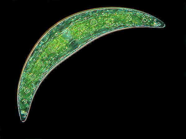
This organism has wonderfully intricate chloroplasts and at each tip, a spherule containing minute granules which are in constant motion.
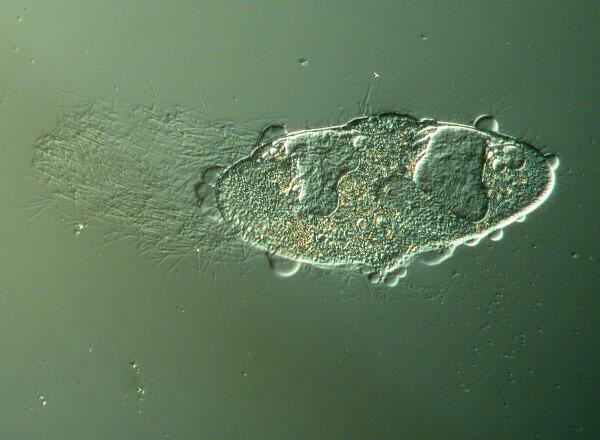
The aforementioned Paramecia are, of course, in evidence and one can quickly and readily produce rich cultures of them by adding a boiled wheat grain to some boiled pond water or some artesian water from your grocery store; they sell it by the gallon for about a dollar. Let the dish sit for 2 or 3 days to develop a good bacterial growth and then inoculate the dish with a few Paramecia. They are amazing organisms which are often treated quite casually because they are so common. In another sense, however, they are quite uncommon, that is, unusual.
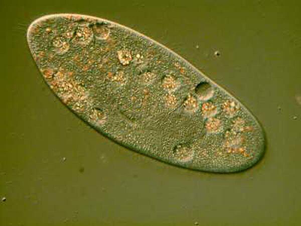
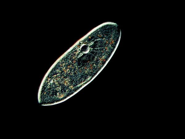
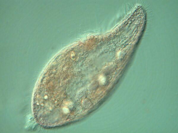
These are images of living Paramecia taken using Nomarski Differential Interference Contrast. This is a wonderful technique for certain kinds of specimens. Its major disadvantage is that this system regards even a Paramecium as being quite thick and so a Nomarski system produces a series of optical sections of “slices” which means that you can use it as a kind of optical microtome without having to kill and embed your specimen. The great advantage, of course, is that you have something rather like Computer Assisted Tomography–a CAT scan–especially if you have some computer stacking software, such as, Helicon Focus which will allow you to take a series of images just altering the focal plane by a few microns each time and then using the software to produce a single combined image. The results can be very impressive.
However, at this point let’s go back to the first Nomarski image of the Paramecium. The detail which is revealed–and this is a single image, not a composite one–is quite remarkable. You can see the macronucleus, the contractile vacuoles, the cilia, the capsules along the edge of the membrane which house the trichocysts, and all sorts of internal granules.
The reason I have been going on at such length about Paramecium is that textbooks often present it as a “typical” ciliate whereas, in fact, it is anything but. Even within the genus, there is a lot of variation. Paramecium caudatum, P. aurelia, and P. multimicronucleatum are perhaps the most common species. P. caudatum has been studied extensively and is often taken as the prototypical species. P. aurelia has also been the focus of intensive research both in terms of aging and mating types. Paramecia present us with a complex behavior which we might very well not anticipate–SEX!
P. aurelia, depending upon the suspecies or variant, divides about 200 to 250 times before it needs recharging of its genetic material by means of conjugation and the exchange of micronuclei with a different mating type. The problem is that we haven’t yet learned how to distinguish mating types, but the Paramecia manage it. When it all goes smoothly, then they can start dividing again and as long as they keep repeating the process, they achieve a kind of theoretical “immortality”.
There is another strain of Paramecium which harbors a genetic fragment know as a “kappa” particle. When it conjugates with a non-kappa strain, some of the kappa particles get transmitted when the micronuclei are exchanged and these particles turn out to be lethal to the non-kappa strain thus allowing the kappa strain to maintain a kind of dominance.
P. multimicronucleatum, as its name suggests, has a large number of micronuclei, whereas other species may have only 2 or 4. The biological advantages of this are not altogether clear. Another species, P. bursaria, possesses symbiotic algae (Chlorella) and their photosynthetic activity helps provide food for the larger organism. However, when the light conditions are poor so that the symbionts can no longer do this, the Paramecium digests them.
Another puzzle about Paramecia is the trichocysts. These are easily demonstrated by adding a drop of dilute tannic acid to the edge of a cover glass on a slide containing Paramecia. The trichocysts are ejected with considerable force and sometimes to an astonishing distance, relatively speaking, yet they don’t seem to do anything
They don’t’ appear to have any offensive or defensive significance and yet they are abundant and require a considerable amount of energy both in their production and in their discharge.
In order to not completely neglect the title of this essay, I am going to devote the rest of it to a small gallery of images with very brief comments and I shall make every effort to avoid going off on tangents (a difficult task for me).
A Mini-Gallery of Some Likely Spring Pond Organisms
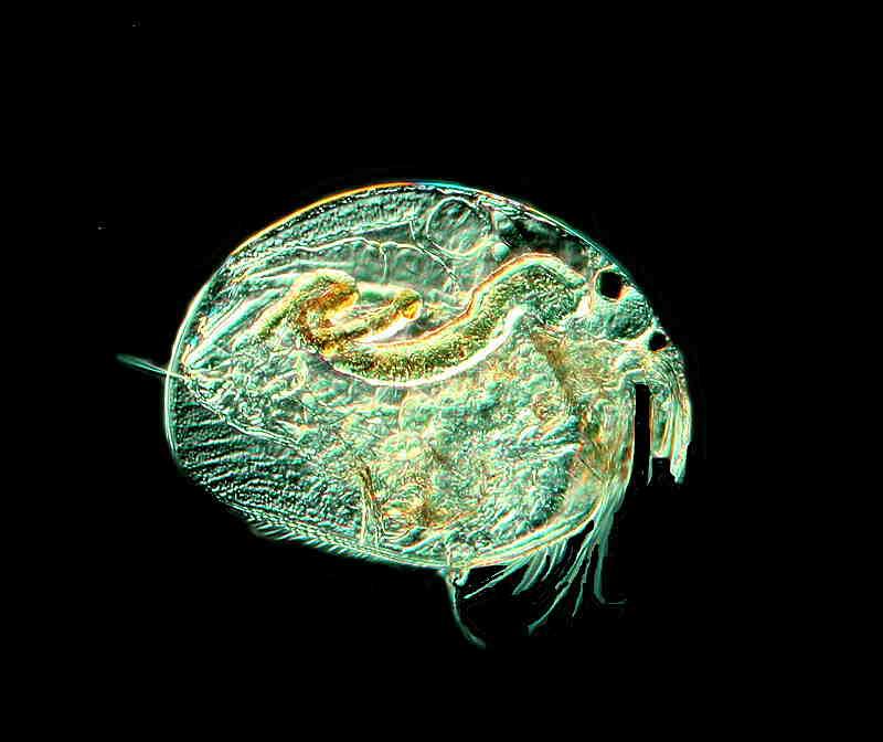
This is a Cladoceran which is a micro-crustacean, related to another wonderful spring creature, the “water flea”, Daphnia.
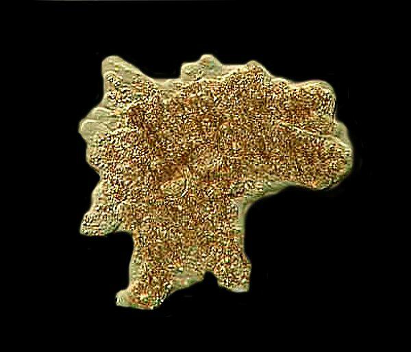
This is Amoeba proteus, one of the largest amoebae, which is surprisingly difficult to find in nature but, fortunately cultures easily.
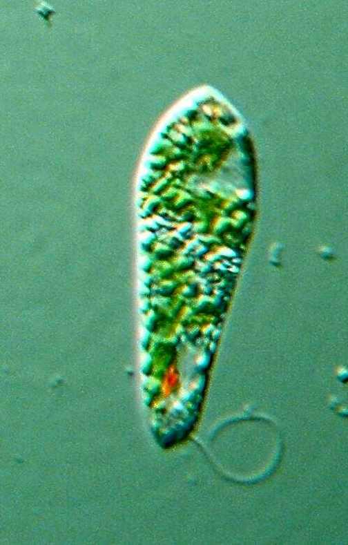
Stentor coeruleus is the marvelous “trumpet-shaped” ciliate with the distinctive and unique dichroic pigment called “stentorin”. In most situations, it presents itself as a blue-green but, in certain angles of illumination, it takes on a rosy-pink hue.
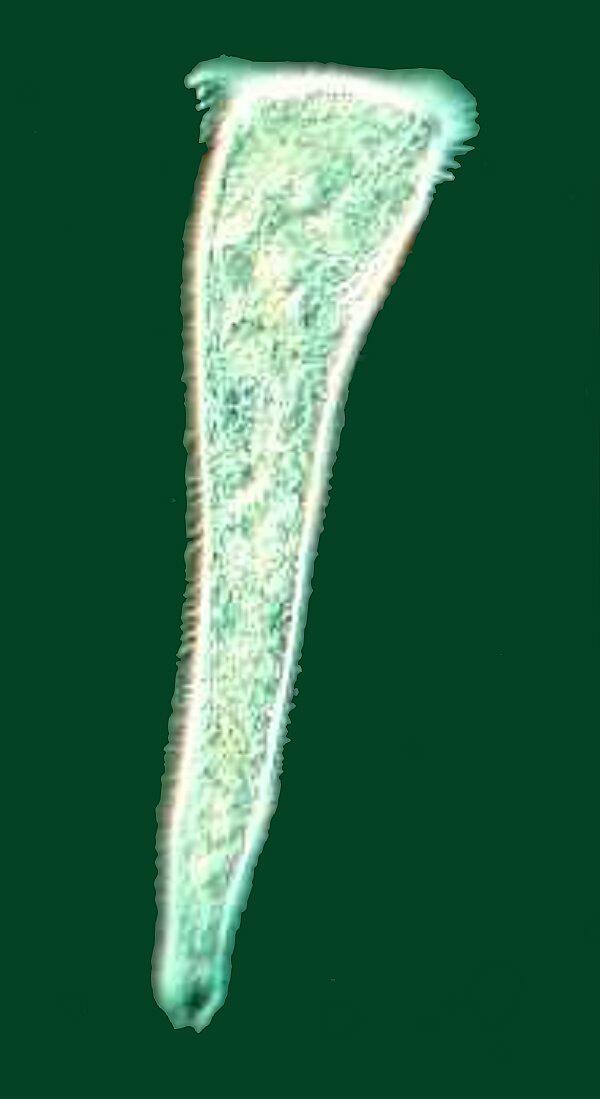
Bursaria truncatella is one of my favorites and whenever I see it, I think of an elegant, animated Steuben crystal vase sliding through the water. Unfortunately, I have not had the best of luck in capturing good images of it, so you’ll have to settle for the one above.
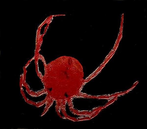
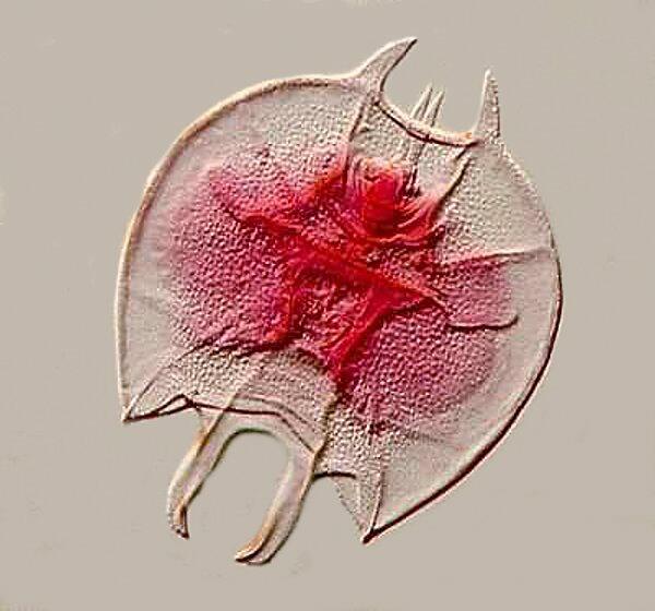
Another group of frequent spring and summer pond denizens is water mites. The one I encounter most frequently is a brilliant scarlet color, but there are others which are brown, orange, light-greenish gray, and some that are brown with yellow spots.
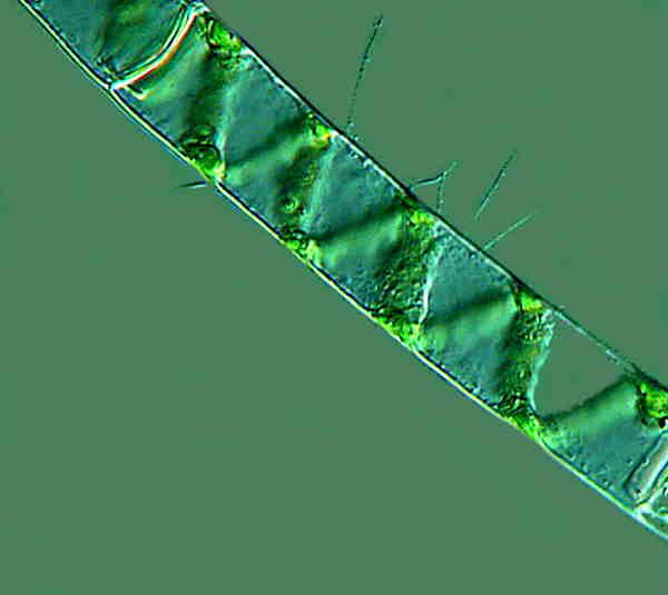
Spirogyra is perhaps the commonest and one of the most delightful of the filamentous algae. Its name is most fitting, for you can see the chloroplasts forming a green spiral in each of the segments. It has a complex life cycle and you should always keep an eye out for conjugating strands.
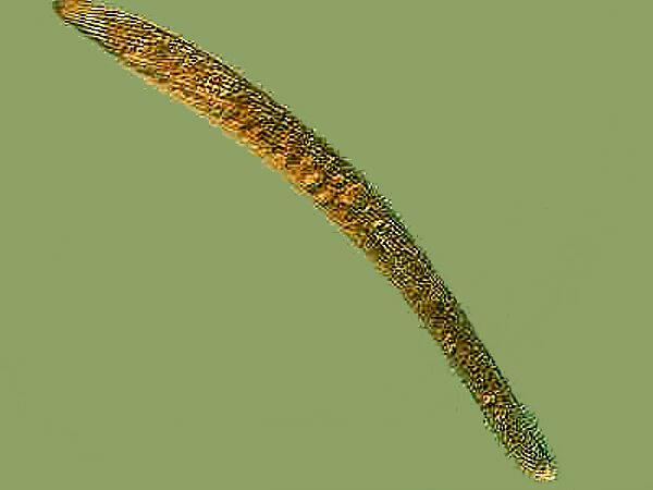
Spirostomum, a giant among ciliates, is also readily found, especially in ponds where cows or wild ruminants come to drink and often leave behind feces. Spirostomum thrives in such environments rich in organic material. S. ambiguum often exceeds 2000 microns in length and is highly contractile. Like Paramecia, the species of Spirostomum vary considerably and some are much smaller and have only a single ovoid macronucleus whereas others have long chain or beaded macronuclei.
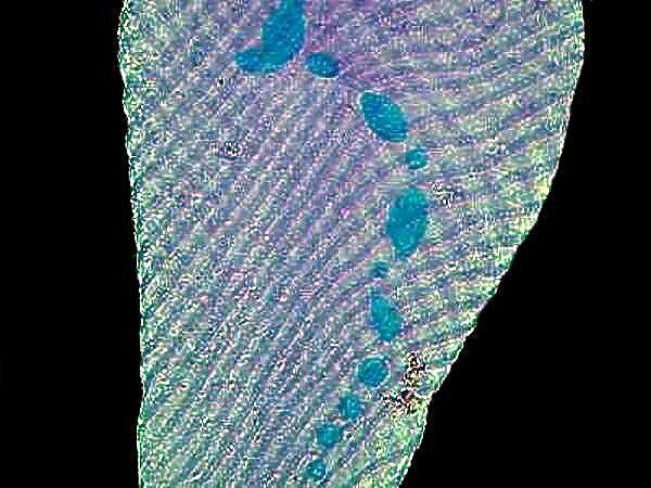
Here is a specimen of S. minus which I stained with Methyl Green Acetic and you can clearly see the long macronucleus in spite of the distortion of the organism.
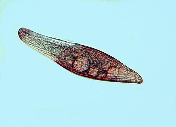
In this specimen of Euglena, you can see the red eye spot, the chloroplasts, and one of the flagella. Euglenoids frequently have a trailing flagellum (or two or three) which are often difficult to observe. This is where phase contrast becomes exceptionally valuable.
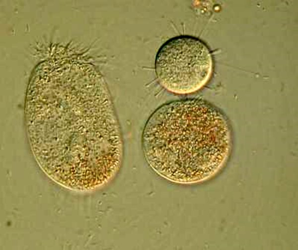
If you are patient and search carefully, you may have the good fortune to come across a suctorian. These are one of the oddest of the ciliates and pose a real challenge for taxonomists. They do have a free-swimming stage which is ciliated, but once they “settle down”, they produce tentacles. You can see that an unfortunate ciliate, larger than the suctorian, has gotten trapped by one of the adhesive pads at the tip of a tentacle and the suctorian is indeed sucking its protoplasm through its tentacle like a straw.
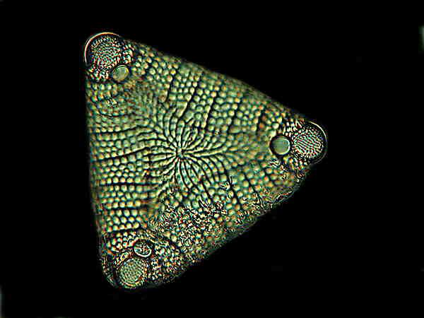
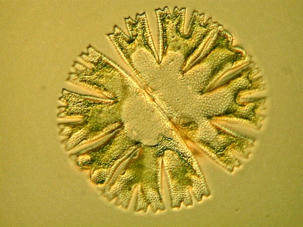
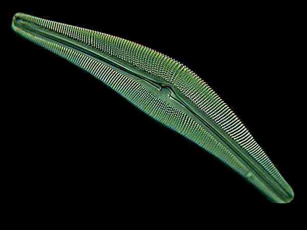
The spring is also a splendid time to collect diatoms which occur in a mind-boggling variety of shapes and sizes. There are two major groupings, the centric and the pennate diatoms. Below, I will show you an example of each.
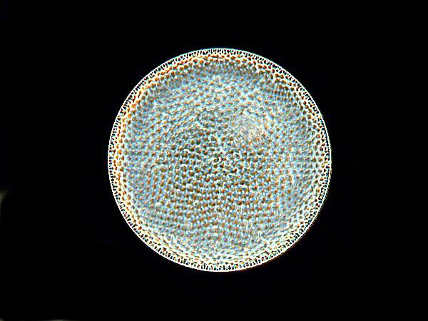
The majority of diatoms occur as single individuals, but quite a few occur as aggregates or shins of individuals and some of these are strikingly beautiful. For nearly 2 centuries, microscopists have been so taken with diatoms that they have made arrangements of them. Below is a modest example of an exhibition slide.
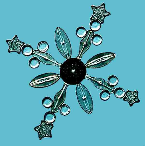
In the 19th Century, J.D. Moeller and his brother produced a diatom exhibition slide containing over 4000 diatoms!
Rotifers also occur in a staggering variety of forms, sizes and shapes. The specimen above is of the genus Keratella and I stained it with a drop of Basic Fuchsin. You can see the sculpting on its protective envelope. This is the rotifer that I think looks rather like a Klingon warship. There are also colonial forms, such as, Conochilus and tube-dwelling forms, some of which, such as Floscularia, construct the tube out of its own fecal pellets.
Another pigmented ciliate which often shows up in samples is Blepharisma, most species of which are pink, although there are some blue ones and some that are albino. They are fairly closely related to Spirostomum, but they do not exhibit the contractility so characteristic of Spirostomum.
These protists could never be presented as examples of a typical ciliate. In addition to possessing a distinctive pigment, they readily form distinctive and unusual cysts, they are voracious feeders and under certain conditions start eating each other and form what are known as “cannibal giants”.
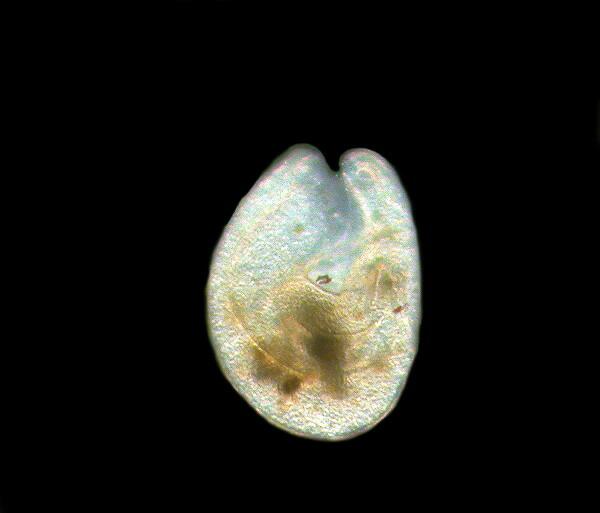
Finally, a creature that I find in abundance in my spring collections is the ostracod also know as “seed shrimp”. I think of them as micro-versions of Pac-Man.
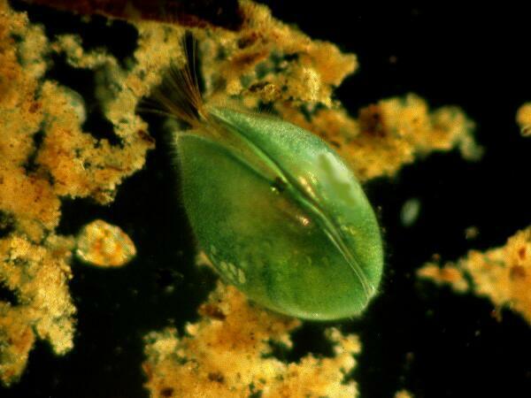
This particular species has a distinctively rich green color They show considerable color variation and the pigment seems to be embedded in the valves of the shell. Some are brown, others light yellow to light orange, some are almost black and others possess very little observable pigmentation. These micro-crustaceans seem to be incessantly scurrying around feeding or seeking food–if you watch them for more than 10 minutes, you’ll feel exhausted.
For me, spring collecting is a special delight after a long winter of snow, wind, and frozen ponds and lakes. Don’t get me wrong, I love the special character of our winters– I could never live in Bagdad or Tucson–but the seasonal explosion of life forms after a winter of hibernation is a glorious thing to experience.
All comments to the author Richard Howey are welcomed.
Editor's note: Visit Richard Howey's new website at http://rhowey.googlepages.com/home where he plans to share aspects of his wide interests.
Microscopy UK Front
Page
Micscape
Magazine
Article
Library
Please report any Web problems or offer general comments to the Micscape Editor .
Micscape is the on-line monthly magazine of the Microscopy UK website at Microscopy-UK .